Augusti D, Augusti G, Re D Undetected excess cement at marginal areas of zirconia crown copings: in vitro analysis of two luting agents and their influence on retention. Int J Prosthodont. 2020; 33:202-211 https://doi.org/10.11607/ijp.6531
Wadhwani C, Rapoport D, La Rosa S Radiographic detection and characteristic patterns of residual excess cement associated with cementretained implant restorations: a clinical report. J Prosthet Dent. 2012; 107:151-157 https://doi.org/10.1016/S0022-3913(12)60046-8
Gaile M, Papia E, Zalite V Resin cement residue removal techniques: in vitro analysis of marginal defects and discoloration intensity using micro-CT and stereomicroscopy. Dent J (Basel). 2022; 10 https://doi.org/10.3390/dj10040055
Morimoto S, Albanesi RB, Sesma N Main Clinical outcomes of feldspathic porcelain and glass-ceramic laminate veneers: a systematic review and metaanalysis of survival and complication rates. Int J Prosthodont. 2016; 29:38-49 https://doi.org/10.11607/ijp.4315
Astasov-Frauenhoffer M, Glauser S, Fischer J Biofilm formation on restorative materials and resin composite cements. Dent Mater. 2018; 34:1702-1709 https://doi.org/10.1016/j.dental.2018.08.300
Pereira S, Anami LC, Pereira CA Bacterial colonization in the marginal region of ceramic restorations: effects of different cement removal methods and polishing. Oper Dent. 2016; 41:642-654 https://doi.org/10.2341/15-206-L
de Brito O, Sandes JM, de Lima F The influence of cement removal techniques on in situ bacterial adhesion and biodegradation at the marginal interface of ceramic laminates. Oper Dent. 2022; 47:190-201 https://doi.org/10.2341/20-269-L
Kurian N, Varghese KG, Wadhwa S A technique to avoid the entrapment of excess residual luting cement in soft-tissue irregularities underneath fixed dental prostheses by using polytetrafluoroethylene tape. J Prosthet Dent. 2023; 130:271-272 https://doi.org/10.1016/j.prosdent.2022.02.010
Magne P, Kim TH, Cascione D, Donovan TE Immediate dentin sealing improves bond strength of indirect restorations. J Prosthet Dent. 2005; 94:511-519 https://doi.org/10.1016/j.prosdent.2005.10.010
A technique for easy removal of excess resin cement Ignacio Farga-Niñoles Mihaela Teris Dental Update 2024 51:2, 707-709.
Authors
IgnacioFarga-Niñoles
DDS, MSc, BDS
Master in Prosthodontics, DipImplantDent RCSEng, MSc ImplantDent, Clinical Teacher and Academic Advisor, Restorative Department, Dental Institute, Queen Mary University of London
In the context of cementing indirect dental restorations, the inadvertent bonding of excess cement to adjacent teeth poses a multifaceted challenge, affecting aesthetics, periodontal health and chair time. Using PTFE tape as the sole method for isolation can be time consuming, and occasionally presents challenges. The approach described in this Technique Tip involves manually holding a matrix holder to safeguard neighbouring teeth from sandblasting and etching contamination, providing a pragmatic resolution to these issues. It seamlessly integrates with the established practice of using PTFE tapes to prevent unwanted bonding and cement attachment to adjacent teeth.
CPD/Clinical Relevance: Combining PTFE and matrix bands optimises bonding of indirect ceramic restorations for periodontal health and efficiency.
Article
Cementing indirect restorations is a procedure that demands a high level of skill and precision, often causing considerable stress. Multiple factors contribute to the potential failure of indirect restorations and the inadvertent damage to neighbouring teeth and soft tissues. One significant factor is the unintended bonding of excess cement to adjacent teeth during the placement of indirect restorations.1 This excess cement can be challenging to detect, cumbersome to eliminate, and particularly time consuming, especially when it adheres to adjacent dental structures.1 Clinicians often require dental radiographs to identify the presence of cement excess, resulting in a patient's exposure to radiation.2
Aesthetics: excess cement can lead to a discernible cement margin on the adjacent tooth, resulting in a poor aesthetic result;3,4
Biofilm adhesion: cement excess adhered to neighbouring teeth can be plaque-retentive and may promote gingival inflammation, periodontal complications and/or carious lesions;5,6
Extended chair-time: excess cement is challenging to remove, especially if bonded to the adjacent structures, leading to longer chair time for both the patient and the dentist.7
Technique
Establish rubber dam isolation during the preparation and cementation of any indirect partial restoration to minimize salivary contamination and prevent any excess restorative material from coming into contact with the patient's soft tissues (Figures 1–3).
Manually secure the matrix retainer to safeguard adjacent teeth during sandblasting and etching, preventing potential damage that may facilitate inadvertent excess cement bonding that could complicate its removal (Figure 3).
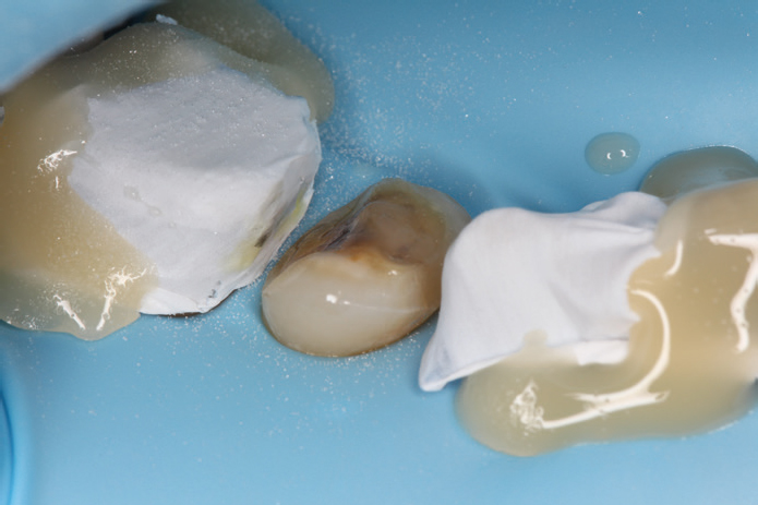
Apply PTFE (polytetrafluoroethylene) tape to shield adjacent teeth from contact with adhesive and cement (Figure 4).
Carefully deliver the indirect restoration onto the prepared tooth (Figure 5).
Remove any excess cement that may have seeped under the PTFE tapes during the cementation process (Figure 6).
Verify the complete removal of any excess cement from various angles (Figures 7–9).
Figure 1. The lower right premolar had a cracked amalgam restoration, temporarily addressed by a glass ionomer cement seal, and a crack line on the mesial proximal ridge.Figure 2. Upon the removal of the amalgam restoration, caries was apparent beneath. With the existence of a crack line on the mesial proximal ridge and the remaining buccal and lingual cusps measuring less than 1.5 mm in thickness, cuspal coverage may be considered necessary.Figure 3. Lower right premolar prepared to receive an indirect cuspal coverage restoration.Figure 4. A matrix retainer is held during sandblasting, followed by orthophosphoric acid.Figure 5. PTFE tape applied to shield the adjacent teeth and prevent contact with the adhesive.Figure 6. Delivered ceramic indirect cuspal coverage restoration.Figure 7. Cement excess that seeped under the PTFE tapes during the cementation process.Figure 8. Confirmation of successful removal of cement excess from an occlusal perspective.Figure 9. Confirmation of successful removal of cement excess from a lateral perspective.
Discussion
PTFE tape has been employed to shield adjacent teeth from orthophosphoric acid contamination and sandblasting.8 Nevertheless, sandblasting using 50-µm aluminum oxide particles at 2 bars of pressure typically results in PTFE tape degradation. As a result, the defective PTFE tapes should be replaced before etching. PTFE tapes are often aspirated when the etching process is meticulously rinsed. The constant replacement of PTFE tapes significantly prolongs and complicates the procedure.
There have been attempts to place metal matrices during the sandblasting and etching without a holder, but the retention of the matrices tends not to be optimal, and they can be easily dislodged, especially on posterior teeth.
The technique described in this Technique Tip simplifies this process. It entails the extra-oral manipulation of a matrix retainer held in one hand. This allows for the retention of the matrix band around the tooth during sandblasting, effectively preventing aluminum oxide particles from affecting neighbouring teeth without the displacement of the matrix band. Furthermore, the same technique can be applied to hold the matrix band against the tooth during the etching procedure to prevent any contamination of adjacent teeth. The matrix band can have varying thicknesses since its primary purpose is to serve as a physical barrier. In case of bonding a partial indirect restoration that does not include all the proximal walls, wedges can be used to displace the tooth laterally, allowing the matrix to enter the area without disrupting the intact contact.
After the etching procedure, the targeted tooth has been appropriately conditioned, whereas the adjacent teeth have not undergone such treatment. This practice is crucial to decrease inadvertent cement bonding to neighbouring surfaces, and reduce the chair time necessary to remove it
Subsequently, PTFE tapes can be secured to the adjacent teeth using non-self-adhesive composite to ensure their stability. The clinician can then proceed with the recommended cementation protocol to place the desired indirect restoration. Upon completion of the cementation process, none of the neighbouring dental structures will have been conditioned, ensuring easy removal of any excess cement.
A limitation inherent to this method is the limited visibility of the complete tooth contour, potentially jeopardizing the use of dentine-friendly etching techniques, such as selective etching. To address this, one effective approach is to avoid overly securing the matrix, affording better visualization. Alternatively, we suggest employing the immediate dentine sealing (IDS) technique during the preparation process. This method effectively seals the dentinal tubules, ensuring that subsequent etching affects only the enamel, without affecting the dentine.9
An additional limitation is related to the growing consumption of disposable materials in dentistry, which is environmentally unsustainable. Consequently, we advise the use of autoclavable matrix retainers, where the sole disposable component is the matrix band itself.
Summary
The use of a matrix holder to shield neighbouring teeth from sandblasting and etching contamination, combined with the use of PTFE tapes to prevent undesired bonding and cement adhesion to adjacent teeth, offers several key benefits:
Excess composite cement is less prone to adhere to the adjacent surfaces, reducing the risk of biofilm accumulation that may contribute to potential carious lesions or periodontal issues.
Reduced chair time enhances both the patient's experience and the efficiency of clinicians.
Simplified cement removal instils confidence in clinicians, enabling them to minimize the need for postoperative radiographs to identify cement extrusion and reduces the patient's radiation exposure.
The lack of cement adhesion on the adjacent teeth will lead to no discernible cement margin, resulting in a better aesthetic result.
Well-designed clinical studies are necessary to draw definitive conclusions about the implementation of this new technique when compared with the use of PTFE alone.