A MEDLINE search early in 2015 revealed more than 250,000 papers on head and neck cancer; over 100,000 on oral cancer; and over 60,000 on mouth cancer. Not all publications contain robust evidence. We endeavour to encapsulate the most important of the latest information and advances now employed in practice, in a form comprehensible to healthcare workers, patients and their carers. This series offers the primary care dental team in particular, an overview of the aetiopathogenesis, prevention, diagnosis and multidisciplinary care of mouth cancer, the functional and psychosocial implications, and minimization of the impact on the quality of life of patient and family.
Clinical Relevance: This article offers the dental team an overview of the use of radiotherapy, and its effects on the mouth and other tissues.
Article
Nicholas Kalavrezos Crispian Scully
Radiotherapy (RT) uses high-energy rays to destroy cancer cells, while trying to minimize harm to normal cells. X-rays were the first form of photon radiation to be used to treat cancer. The higher the energy of the x-ray beam, the deeper x-rays penetrate the target. The daily radiation dose must be enough to destroy cancer cells while minimizing damage to normal tissues: typically 2 Gy is delivered daily to a 64–70 Gy total dose (Gray [Gy] = energy absorption of 1 joule/kg [1 Gy = 100 rads]).
Radiotherapy treatment (RT)
Radiotherapy alone is used to treat some types of mouth and oropharyngeal cancers. RT is an extremely effective treatment for oral squamous cell carcinoma (OSCC), sometimes as a primary modality or as an adjuvant following surgery. RT may be used as the sole treatment modality for primary oral cancers without obvious lymph node involvement, for base of tongue cancers and also for inoperable tumours. Even if surgery is the main treatment, radiotherapy may still be recommended after surgery to ablate any residual cancer cells, lowering the risk of recurrences. Radiotherapy complicates any further surgery because, in particular, the radiation endarteritis impedes tissue healing, predisposing to osteoradionecrosis. Radiotherapy may also be used palliatively to shrink a tumour or stop bleeding from it. It can also relieve symptoms if a cancer has metastasized elsewhere.
Radiotherapy may be combined with chemotherapy for treating locally advanced cancer (Article 12).
Forms of radiotherapy
Radiotherapy can be given either as:
External Beam RadioTherapy (EBRT) or plesiotherapy, the most common modality. A beam of x-rays or electrons is directed at the cancer from a linear accelerator;
Internal radiotherapy, also called interstitial radiotherapy or brachytherapy which involves a radioactive source inserted into the tumour itself.
Radiotherapy, to be as effective as possible with minimal adverse effects, must be carefully planned by the clinical oncologist. This may take a few visits. On the first visit to the radiotherapy department, a CT or MRI of the area to be treated is taken, and radiographers take measurements for the radiotherapy planning computer. Occasionally, the skin may need to be marked to help radiographers position the patient accurately before each treatment. To enable this, an immobilization shell (a transparent plastic positioning mask) is placed to hold the head and neck still for each radiotherapy session. This mask is needed for up to about 15 minutes at a time.
Radiotherapy only takes a few minutes at each session but treatment extends over 3–7 weeks, depending on the type and size of the cancer. Treatment is given in the hospital radiotherapy department and can be planned in different ways:
Monday–Friday, with a rest at the weekend (the most common method);
More than once a day;
Every day including at the weekend.
If the radiotherapy is likely to cause swelling around and embarrass the airways, a temporary tracheostomy may need to be arranged.
External beam radiotherapy
Combining computerized tomography (CT) with fluorodeoxyglucose positron emission tomography (FDG-PET) imaging pre-treatment helps in significantly better defining tumour outlines and facilitates potentially different radiotherapy options compared to using CT alone.
The main types of EBRT used include the following.
Conformal RadioTherapy (CRT)
This is the most common type of EBRT used for head and neck cancers. Shaping the radiotherapy beams reduces radiation to surrounding healthy cells.
Intensity Modulated RadioTherapy (IMRT)
IMRT uses 3-D computerized treatment planning and conformal therapy with x-ray accelerators to deliver radical RT to the target while sparing most normal tissue, thereby reducing toxicities and improving outcomes in patients with OSCC, and their quality of life (QoL). IMRT irradiates irregularly-shaped volumes and can produce concavities in treatment volumes, thus permitting more precision and greater sparing of normal structures including:
Salivary glands, showing a significant reduction in hyposalivation;
Cochlea, with potential to reduce radiation-induced hearing loss;
Optic nerves, brain stem, and spinal cord.
Volumated intensity Modulated Arc Therapy (VMAT)
VMAT delivers IMRT-like distributions in a single rotation of the gantry (eg RapidArc) potentially offering shorter planning and treatment time, better dose homogeneity and better sparing of normal tissue.
Image Guided RadioTherapy (IGRT)
IGRT uses adaptive RT based on regular scanning and planning to reduce dosimetric uncertainties associated with the volume changes in tumours and organs at risk. Positron Emission Tomography (PET) using two tracers (fluorine-18-labelled fluoromisonidazole (F-MISO) and copper (II)-diacetyl-bis(N(4)-methylthiosemicarbazone (Cu-ATSM) can highlight hypoxic areas where the RT dose can be escalated without additional acute toxicity.
Intensity Modulated Proton Therapy (IMPT)
IMPT permits 3-D dose distributions using charged particles like protons which deposit little energy until they reach the end of their range, when most of their energy is deposited in a small area. The advantages are of low radiation dose to normal tissue with tissue sparing, and better dose homogeneity.
Internal radiotherapy
Putting a radioactive source directly into the cancer gives a high dose of radiotherapy into the tumour in order to:
Treat small tumours in the mouth or on the lip;
Use with EBRT to give an additional radiotherapy or ‘boost’ into the tumour.
Implants, usually of iridium (Ir192) for a few days are often used for brachytherapy, supplying a radiation dose equivalent to teletherapy but confined to the lesion and immediate area. There are three main types of brachytherapy:
High-Dose Rate (HDR) brachytherapy;
Pulsed-Dose Rate (PDR) brachytherapy;
Low-Dose Rate (LDR) brachytherapy.
High-Dose Rate (HDR) brachytherapy
High-dose rate brachytherapy is a fast, precise treatment in which laser-thin, hollow catheters deliver a precise, three-dimensional dose of radiation into a tumour under image-guidance (ultrasound, CT scan or MRI), delivering a maximum dose with minimal exposure to the surrounding healthy tissue. Each HDR brachytherapy treatment takes 15–20 minutes and, after a treatment series, the catheters are removed leaving no radioactive seeds.
Pulsed-Dose Rate (PDR) brachytherapy
Pulsed-dose rate brachytherapy is similar to HDR, but radiation from iridium192 is delivered in short ‘pulses’ over several hours.
Low-Dose Rate (LDR) brachytherapy
Low-dose rate brachytherapy can be:
Temporary: a thin, hollow, plastic tube is inserted in and around the tumour loaded with radioactive seeds which remain in place for a few days. This requires a hospital stay, and visits from family, nurses and other caregivers are limited as a precaution. After a few days, the seeds and tubes are removed, with no radiation remaining.
Permanent: radioactive seeds are inserted directly into the tumour and left permanently in place.
With internal radiotherapy, there may be some swelling in the tissues nearby and the patient must remain alone in a single hospital room for a few days, until the oncologist removes the radioactive source. During this time, all visitors including staff are very restricted and visits by children and pregnant women are forbidden. Once the radioactive source is removed it is perfectly safe to mix with other people.
What other care is required before radiotherapy?
Smoking during radiotherapy reduces its effect and is likely to increase adverse effects.
Oral healthcare
Prevention and treatment of oral complications whenever possible are important and should be performed by a dental practitioner and oral hygienist. Regular mouth care is crucial during and after radiotherapy. Mouthwashes and protective gels may help. Alcohol, especially spirits, will irritate the areas affected by treatment. The patient should:
Brush teeth (and appliances) with a small, soft toothbrush after each meal;
Rinse with a non-alcohol based mouthwash;
Clean daily between teeth with dental floss or tape;
Use a fluoride toothpaste and fluoride gel or mouthwash daily;
Sip plain tapwater often and rinse mouth regularly;
Avoid sugary foods or drinks between meals;
Avoid acidic drinks, such as cola, carbonated drinks and fruit juices;
Exercise the jaw, to prevent trismus;
Visit the dentist and hygienist every 3–6 months.
What complications can be anticipated after radiotherapy?
Radiotherapy to the head and neck can cause temporary adverse effects such as a sore skin, mouth or throat and difficulty swallowing; these side-effects are usually more severe with chemo-radiotherapy. Longer-term oral complications may include dry mouth (and sequelae), loss of taste, osteoradionecrosis (ORN), trismus and other problems. Radiotherapy has, however, improved over the years with advances in imaging, treatment planning computer software and in radiation delivery technology.
Adverse effects usually begin to appear during the second week of radiotherapy and may continue to get worse for 7–10 days after treatment ends, before gradually improving. Most patients find that adverse effects have noticeably improved 6–8 weeks after radiotherapy has ended but sometimes they can develop months or even years later, especially dry mouth.
Mucositis, dysphagia, xerostomia, dermatitis and pain significantly impair QoL, as do hyposalivation (up to 90% incidence) and grade 3 (severe) dysphagia (up to 30%). Late RT toxicity is permanent and may also include:
Osteoradionecrosis of the jaw (ORN);
Sensori-neural hearing loss;
Skin fibrosis;
Laryngeal cartilage necrosis;
Cervical atherosclerosis.
Mucositis
Radiotherapy is most often administered in small fractions over several weeks and to a localized area. Radiation-induced mucositis is invariable within the radiated field of mucosa and typically begins at cumulative doses of about 15 Gy (ie after around 10 days) and reaches full severity at 30 Gy, persisting for weeks or months (Table 1). Risk factors for radiation mucositis are the radiation dose and fractionation but also include:
Alcohol use;
Concurrent chemotherapy;
Dental disease;
Poor oral hygiene;
Younger age.
Week following radiotherapy
1
2+
3+
Later
NauseaVomiting
MucositisTaste disturbance
Dry mouth
CariesInfectionsOsteoradionecrosisPulp pain and necrosisTooth hypersensitivityTrismus
Tissues such as the soft palate, and the lateral borders and ventral surface of the tongue and floor of the mouth, which have a good vascular supply or a higher cell turnover rate, are the most susceptible to radiation mucositis.
Mucositis can lead to a number of problems, including:
Acting as a portal for septicaemia, especially streptococcal, sometimes with lethal consequences;
Extending hospitalization;
Increasing costs of care;
Significantly interfering with QoL.
Mucositis can be reduced by:
Minimizing doses and field of radiation;
Using mucosa-sparing blocks;
Using amifostine before therapy;
Avoiding chemo-radiotherapy;
Betamethasone mouthwashes.
The time to healing of mucositis depends on the radiation dose intensity, but is usually complete within 3 weeks after the end of treatment. Tobacco smoking delays resolution.
Management includes:
Avoiding irritants (smoking, spirits or spicy foods);
Good oral hygiene;
Combating pain and dysphagia with
- opioids, such as morphine and hydromorphone;
- topical analgesics used prior to meals, such as:
- benzydamine hydrochloride;
- lidocaine (lignocaine mouthwash;
- aspirin.
There are many other preparations used, often variants on the ‘magic mouthwash’ (viscous lidocaine, diphenhydramine, bismuth salicylate and a corticosteroid).
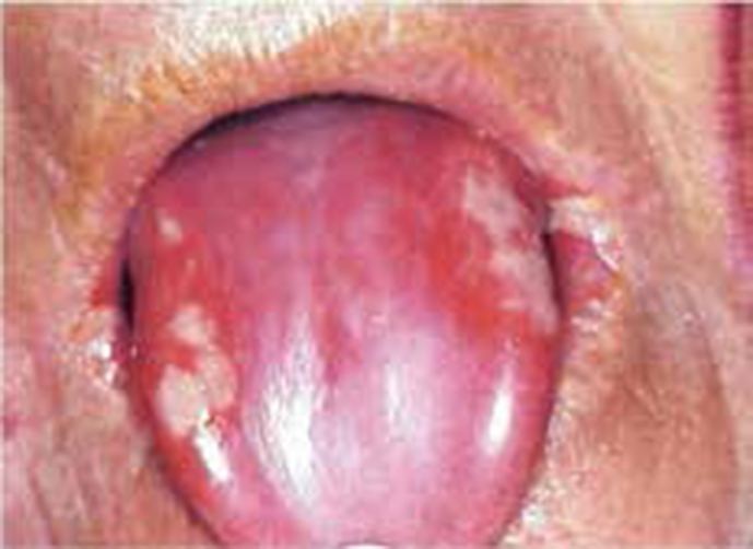
Mucositis begins to appear at approximately 10–12 days after the start of irradiation, but usually heals within 3 weeks after the end of treatment (Figure 1).
Figure 1. Radiation mucositis with erosions, and depapillated tongue.
Tobacco smoking delays resolution. Mucositis pain can be intense and often interferes with eating and quality of life; opioids may be needed for analgesia. In addition, mucositis can provide a portal for microbial entry and thus can result in local and, sometimes systemic, infection.
Preventing or ameliorating mucositis may be possible not only by minimizing exposure to radiation, but also by taking active measures:
Oral hygiene should be maintained;
Advise the patient to eat a soft, bland diet and avoid irritants such as smoking, spirits or spicy foods;
Topical analgesics (eg aspirin, benzydamine, lidocaine, dyclonine or diphenhydramine) may provide relief;
Topical chlorhexidine gluconate and sucralfate may ameliorate mucositis.
High-calorie drinks such as Complan® and Build up® and supplement drinks, such as Ensure® or Fortisip®, may be advised by the dietitian. Oral malodour (halitosis) is usually caused by changes to the saliva, or infection, and can be minimized by regular mouth care.
Salivary tissue is vulnerable to radiation damage, and the parotid glands are most readily damaged. The degree of hyposalivation depends on the degree of exposure of the salivary tissue to RT. Hyposalivation occurs when the upper border of the radiation field is above the submental area, particularly when the parotid glands are involved. Radiotherapy to the nasopharynx damages both parotid glands and causes severe and permanent hyposalivation. Radiotherapy fields used in the treatment of mouth cancer normally try to avoid at least part of the parotid glands; therefore, hyposalivation tends not to be as severe as it would be if both glands were irradiated in their entirety. With conventional radiotherapy for mouth cancer (60–70 Gy), there is a rapid decrease in saliva flow during the first week of radiotherapy, with an eventual 95% reduction. Mantle, unilateral and bilateral fields of radiation can cause a reduction in salivary flow of 30–40%, 50–60%, and 80%, respectively. A single radiation dose, as small as 20 Gy, can cause permanent cessation of salivary flow and, with the conventional RT for mouth cancer (60–70 Gy), there follows a rapid decrease in flow during the first week of radiotherapy, with an eventual approximate 95% reduction. The sensation of dryness of the mouth after RT tends to diminish after a few months to a year, partly because of compensatory hypertrophy of unirradiated salivary glandular tissue. After one year, however, there is rarely little further improvement.
The use of pilocarpine during radiotherapy has shown encouraging results in minimizing dryness. In addition to minimizing unnecessary glandular irradiation, stimulating the salivary glands with pilocarpine prior to and during radiotherapy may reduce glandular damage.
Hyposalivation predisposes to caries, candidosis and bacterial acute ascending sialadenitis. Changes may be:
Hyposalivation leads to mouth discomfort and loss of taste and appetite, and may impede speech and swallowing. In addition, it predisposes to infections (caries, candidosis and sialadenitis). Rinsing the mouth regularly with a mouthrinse with half a teaspoon of salt and half a teaspoon of baking soda mixed into a litre of water may help. Xerostomic patients should be advised to avoid agents such as medications, tobacco and alcohol that may further impair salivation. Soft, moist foods with gravies and sauces will be easier to eat than dry or chewy foods. Some find that using a humidifier in the home helps as it makes the atmosphere less dry. Lips can also feel dry and chapped so a lip balm may help, but avoid products that are coloured, perfumed or flavoured.
Residual salivary tissue may be stimulated by gustatory or pharmacologic stimuli (sialogogues). Drugs that may be particularly effective include various cholinergic agents, such as pilocarpine, given as ophthalmic drops placed intra-orally or as tablets. This is effective in relieving symptoms and in improving salivation when used in doses of up to 5 mg administered 3 times daily. Sugar-free chewing gum may be a useful stimulus, is inexpensive, and has no adverse effects. Saliva substitutes or mouth-wetting agents can be helpful; most contain carboxymethylcellulose, although some contain animal mucins and some also contain constituents that may facilitate enamel remineralization (eg amorphous calcium phosphate). Individuals with dry mouth should frequently sip water, particularly during eating, and keep water by their bedsides. Prevention of dental problems is necessary.
Caries
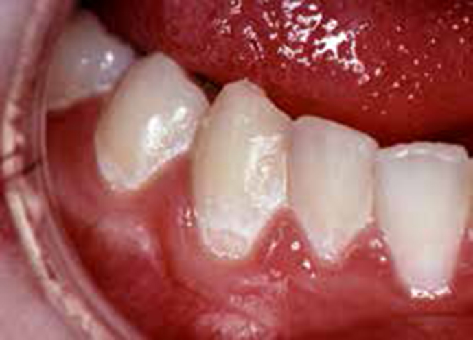
Caries, a bacterial infection associated especially with Streptococcus mutans, is increased after irradiation, can rapidly progress, and affect areas usually not predisposed, such as incisal edges, smooth surfaces, and the lower incisor, as well as other areas. Several types of carious lesions have been identified (Figure 2), most involving the incisal edges and cervical areas. Radiotherapy predisposes to dental caries mainly because of hyposalivation, adoption of foods with a high sucrose content and, possibly, a shift to more cariogenic oral microflora.
Figure 2. Cervical caries after radiotherapy.
Patients should be encouraged to achieve a good level of oral hygiene, avoid foods with a high sucrose content, and use topical fluorides daily for life. Fluoride is applied best to the entire surface of all teeth to have the maximal protective effect. This can be achieved best by providing custom-built carriers for each patient. A gel containing 1% sodium fluoride is put into the carrier and applied to the teeth for 5 min/day. Fluoride mouthrinses are also useful: sodium fluoride rinses with chlorhexidine diacetate may be particularly effective. Amorphous calcium phosphate (ACP) preparations are also caries-protective.
Candidosis
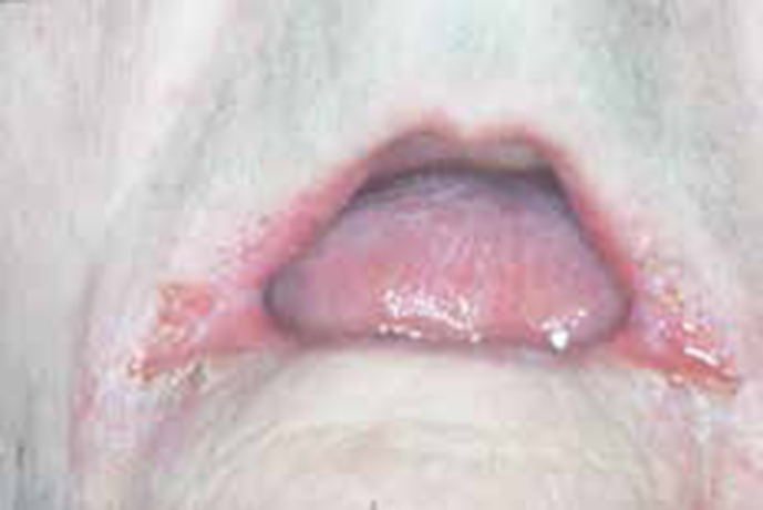
Hyposalivation, dental prostheses, alcohol use and tobacco smoking predispose patients to oral candidosis (Figure 3). A meta-analysis has shown the prophylactic value of topical clotrimazole or fluconazole.
Figure 3. Candiosis (angular stomatitis) after radiotherapy.
Candidosis is an issue for oral cancer patients because of hyposalivation, treatment-related immunosuppression, frequent use of antibiotics and sometimes of cytotoxic drugs. The diet may also favour Candida colonization, if fermentable carbohydrates are frequently consumed. The wearing of dental prostheses or obturators also predisposes.
Sialadenitis
Sialadenitis may follow irradiation and/or chemotherapy and, in turn, may cause irreversible hyposalivation which, with poor general health, renders cancer patients liable to ascending infective (bacterial) sialadenitis, mainly involving Streptococcus viridans and Staphylococcus aureus (often penicillin-resistant), ascending from the oral cavity.
The management of sialadenitis often means hospitalization of the patient and includes:
Analgesia;
Antimicrobials: prompt treatment with amoxicillin (flucloxacillin or amoxicillin/clavulanate if staphylococcal and the patient is not allergic to penicillin; erythromycin or azithromycin in penicillin allergy);
Surgical drainage if there is fluctuation;
Hydration;
Salivation stimulation by use of chewing gum or sialogogues.
Other infections
Oral infections with viruses, bacteria and fungi may significantly increase, particularly after radiochemotherapy. Aciclovir remains the primary treatment for herpes simplex virus (HSV) and herpes varicella-zoster virus infections but new agents, such as famciclovir, penciclovir, sorivudine, foscarnet and other agents, may be needed in cases of aciclovir resistance.
Osteoradionecrosis (ORN)
Radiotherapy causes endarteritis with thrombosis of small blood vessels, fibrosis of the periosteum and mucosa, and damage to osteocytes, osteoblasts, and fibroblasts slowing bone remodelling, eventually resulting in bone thinning and reduced strength. Many damaged osteoclasts and osteoblasts survive until they attempt to divide, for example when stimulated by trauma, at which time mitotic death occurs, and osteoradionecrosis may supervene. ORN is exposed bone in an irradiated mouth, with or without external sinuses, pain and pathological fracture. ORN is defined as exposed irradiated bone tissue that fails to heal over a period of 3 months without a residual or recurrent tumour.
The pathogenesis of ORN is not completely understood but is the result of hypoxic, hypovascular and hypocellular tissue, followed by tissue breakdown leading to a non-healing wound. There is no bone infection, rather only superficial contamination, but surface bacteria may play a fundamental role in the pathogenesis: teeth present in the field of irradiation might represent the port of entry for micro-organisms.
ORN is potentially the most serious oral complication of radiation therapy: 5–15% of patients who undergo radiotherapy to the head and neck region develop ORN. The mandible consists of more compact bone with a higher density than the maxilla; therefore, it absorbs more radiation than the maxilla and is more predisposed to ORN (Figure 4). Risk factors for ORN include:
Radiation related risk factors: total dose, photon energy, brachytherapy, field size, fractionation. With IMRT, only small partial volumes of the jaw bone are exposed to high radiation doses, so this may translate into a reduction of ORN.
Trauma and surgery are the other main risk factors. The risk is increased when tooth extractions are performed after irradiation, but there appears little increased risk of ORN when extractions are performed before radiotherapy. The single most important factor associated with ORN development is mandibular surgery in an irradiated jaw. However, about 50% of ORN are ‘spontaneous’ and appear without a history of previous tooth removal.
Drug use: alcohol and tobacco are risk factors for ORN. In contrast, corticosteroids or anticoagulants used before or after RT reduce the risk of ORN.
Genetics: the development of ORN may be related to the presence of the T variant allele at -509 within the Transforming Growth Factor (TGF-β1) gene.
Figure 4. Osteoradionecrosis.
Various factors predispose to ORN, but the risk is greatest when:
Teeth are extracted after radiotherapy;
Alcohol and tobacco or bisphosphonates are used;
Nutritional status and oral hygiene are poor;
In the mandible;
Higher and frequent radiation doses are used.
Prevention of ORN
Osteoradionecrosis is unlikely with radiation doses below 60 Gy; in doses up to 70 Gy, the rate is 1.8%, and in doses higher than 70 Gy, the rate is approximately 9%. Nowadays, 5–15% of patients who undergo RT to the head and neck region develop ORN. Radiation shields decrease the radiation dose received by the bone and minimize the risk of ORN.
ORN is three times higher in dentate than in edentulous patients, and this has led to a strategy of preventive extractions of all decayed and periodontally compromised teeth before jaw radiotherapy. However, as caries and periodontal disease are so common, controversy has existed regarding whether such teeth should always be removed. Patients about to be treated with RT do need intensive preventive dental treatment but it is now generally accepted that not all teeth with caries and periodontal disease in the high-dose irradiation field need to be extracted. The only teeth that really need to be extracted before RT are those within the high-dose field and that are unrestorable or have advanced endodontal or periodontal involvement. Tooth extractions must be done before radiation therapy, and patients who require multiple dental extractions or extensive surgical extractions, or both, can be given eight weeks of pentoxifylline 400 mg twice daily with tocopherol 1000 IU, starting a week before the procedure, as prophylaxis for ORN. Dental extractions typically are best performed judiciously and a minimum of 2–3 weeks before commencement of irradiation therapy. All other teeth should be cleaned and restored before radiotherapy begins.
If surgery later becomes necessary, irradiated tissue should be handled as gently as possible.
Management of ORN
The treatment of ORN is largely conservative since up to approximately 60% of cases resolve themselves. Therapeutic approaches include local wound care, topical or systemic antibiotics, hyperbaric oxygen (HBO), ultrasound, and minor-to-extended surgery with reconstruction procedures.
Meticulous oral hygiene is essential, including the use of 0.02% aqueous chlorhexidine mouthwashes after meals. Irrigate away debris and allow sequestra to separate spontaneously because surgical interference only encourages extension of the necrotic process. Any sequestrum that loosens should be removed gently, along with any sharp edges of spicules of bone. Antimicrobials are not especially effective because the affected tissues are avascular; months of treatment are necessary. Tetracyclines can be the most useful because of selective bone uptake, and a regimen of 250 mg of tetracycline 4 times a day for 10 days, followed by 250 mg twice daily continued for several months, has been recommended. Add metronidazole at 200 mg 3 times a day in cases of severe infection or when anaerobes are implicated.
Clindamycin is an alternative but a high percentage of infections are caused by clindamycin-resistant micro-organisms, thus beta-lactams should be the antibiotic of choice. In penicillin-allergic cases, a new fluorquinolone, such as ciprofloxacin, probably in combination with rifampin and/or clindamycin, could be an alternative.
Pentoxifylline (PTX), an antioxidant methylxanthine derivative with an anti-tumour necrosis factor α effect, administered for 6 months, or a combination of PTX and alpha tocopherol (vitamin E), another antioxidant, significantly accelerates healing. The addition of sodium clodronate has been also suggested (pentoxifylline-tocopherolclodronate combination: PENTOCLO).
Hyperbaric oxygen (HBO) therapy has been suggested but is controversial. HBO has been advocated for pre-operative and post-operative treatment of ORN in high-risk patients having teeth extracted or other operations. However, HBO without aggressive surgical management is inadequate, and a random, placebo-controlled, double-blind study failed to show any benefits of HBO over placebo in recovery, in slowing the progression of ORN, or in relieving pain. Adverse effects with HBO therapy are uncommon but include transient myopia, seizures, and otic or pulmonary barotrauma; the latter potentially results in air embolism. Concern has been expressed that HBO therapy may exacerbate a variety of autoimmune and immunosuppressive disorders and viraemia, although little evidence supports this concern.
Untreated pneumothorax is the only absolute contra-indication to HBO. Relative contra-indications include upper respiratory tract infection, chronic sinusitis, epilepsy, chronic obstructive airways disease, high fever, a history of spontaneous pneumothorax or thoracic or ear surgery, viral infections, congenital spherocytosis, and a history of optic neuritis. Risks of HBO therapy may be minimized by a careful pretreatment assessment including chest radiography and electrocardiography. Some advise otolaryngologic and ophthalmologic assessment. Therapeutic ultrasound at a frequency of 3 MHz pulsed 1 in 4 at an intensity of 1 W/cm2 applied for 10 minutes daily for 50 days may also effectively improve ORN.
Surgical management also has played a role in the treatment of ORN and may include sequestrectomy, alveolectomy with primary closure, closure of orocutaneous fistulae, or hemimandibulectomy. The development of myocutaneous flaps and microvascular free bone flaps allows for restoration of mandibular continuity and also brings non-irradiated soft tissue coverage with intact blood supply.
Taste
Radiotherapy to the mouth invariably results in some taste disturbance or loss. Taste receptor cells are relatively radioresistant, and so the mechanism of this taste change has not been elucidated. Hyposalivation possibly contributes because disturbance of taste is particularly common after irradiation of the parotid glands.
Taste disturbance can be distressing and contributes to poor nutrition. Fortunately, taste perception usually recovers slowly within a few months, although sometimes loss is permanent. Zinc sulphate may, in some patients, help improve taste sensation.
Trismus
Radiotherapy that affects the TMJ, or masticatory muscles, may cause trismus, usually because of reduced vascularity from the radiotherapy-induced endarteritis obliterans causing fibrosis in muscles of mastication and surrounding tissues. Tumours needing this type of radiation include nasopharyngeal, base of tongue, salivary gland, and cancers of the maxilla or mandible. Trismus is most likely when the RT is in excess of 60 Gy, and when the patient has been previously irradiated. Radiation-induced trismus may begin toward the end of RT, or at any time during the subsequent 12 months, and tends to increase slowly over several weeks or months. Up to 55% of patients develop trismus in the first 3–12 months after radiotherapy, with continuing loss of oral opening for 24–48 months. Trismus may also result from scar tissue, nerve damage, tumour infiltration or a combination of factors.
Trismus caused by radiation can be difficult to treat, so preventing the development of fibrosis by jaw-stretching exercises is imperative for long-term functionality and QoL. Gentle mouth-opening exercises can help and there are specialist aids available to help exercise the jaw (eg Therabite).
What other post-radiotherapy sequelae can be anticipated?
Fatigue
Fatigue is common during radiotherapy: rest is advised.
Hearing
Radiotherapy for nasopharyngeal cancer may affect hearing, especially if cisplatin is also used. Hearing changes may be temporary and recover after treatment, but sometimes long-term changes develop about 6–12 months after treatment.
Nausea
Sickness is more likely to affect people who have chemo-radiotherapy; anti-emetics can help.
Skin
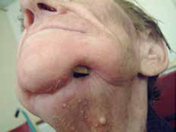
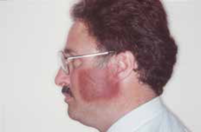
After about two weeks of radiotherapy the skin over the face and neck will usually redden or darken like sunburn, and become sore (Figure 5).
Figure 5. Skin burn from radiotherapy.
This may last for 2–4 weeks after treatment has finished. It is best to use only the soaps, creams and lotions that the MDT recommend, as chemicals can increase sensitivity to radiation. It is best to avoid clothes with stiff or tight collars: loose cotton clothing is better. Do not use sun protection creams on the head and neck during radiotherapy. The skin in the treated area will be more sensitive to the sun during and after radiotherapy (especially the first year): a sun hat and a soft cotton or silk scarf around the neck will help protect the skin.
Thyroid hormones
EBRT to the neck may induce hypothyroidism in 30–40% of patients.
Voice
The voice often hoarsens during radiotherapy but will usually recover after a few weeks. It is best to restrict voice use and avoid smoky atmospheres. A speech and language therapist can give advice.
Summary
Advantages of radiotherapy
Advantages of radiotherapy include that:
Normal anatomy is maintained;
General anaesthesia is not needed; and
Salvage surgery may be possible if radiotherapy fails when it has been applied as the prime treatment modality.
Disadvantages of radiotherapy
Disadvantages mainly include the facts that:
Adverse effects are common;
Cure is uncommon, especially for large tumours; and
Subsequent surgery is more difficult and hazardous and survival is reduced further.