
Article
There are several conditions that affect the extent of mouth opening in the general population, such as oral submucous fibrosis, scleroderma, radiation therapy or surgically induced microstomia.1 Oral submucous fibrosis is a pre-cancerous condition in which palpable fibrotic bands are seen involving the buccal mucosa, floor of the mouth, lips and tongue that causes blanching and stiffening of the oral mucosa leading to trismus.2 In particular, the prosthetic rehabilitation of such patients is further complicated by tongue rigidity, difficulty in chewing, swallowing and speaking. This results in unstable and non-retentive dentures causing pain, discomfort and an array of functional problems (Figure 1).

Many of these apparent clinical difficulties associated with restricted mouth opening can be overcome by sectional impression techniques and modified prosthesis designs such as sectional complete dentures using a Co-Cr framework with hinges or swing-lock attachments, commercially available magnetic attachment systems and flexible dentures.3,4,5 McCord et al used a removable stainless steel post that inserted into three tubes within the complete denture palate to join the two halves of a sectional complete denture.6 Al-Hadi and Abbas incorporated acrylic resin connections in the form of a dovetail with special directions to orient and secure the sectioned mandibular complete denture.7 An alternative method is to use a foldable complete denture through fabrication of a custom-made hinge mechanism using an orthodontic bracket with a buccal tube and a 0.7 mm stainless steel wire.
Procedure
The mandibular primary cast was prepared using putty elastomeric impression material (Aquasil, Dentsply, India) directly adapted over the ridge, as it was impossible to insert a standard impression tray due to greater mandibular arch width and restricted mouth opening.
The mandibular sectional custom tray was fabricated on the primary cast with autopolymerized acrylic resin using a single dowel pin with a special plastic sleeve incorporated into the handle of the tray. The sectional border molding was performed with low fusing compound (DPI Pinnacle, India). A secondary impression was made in two sections with zinc-oxide impression paste (Cavex Outline, Holland) and thereafter reassembled outside the mouth (Figures 2 and 3).


The sectional mandibular trial denture base was made with autopolymerized acrylic resin incorporating press buttons (Snap fasteners, Needles IND) on the lingual flange. Jaw relations were recorded and try-in was performed with the mandibular sectional trial denture (Figure 4).

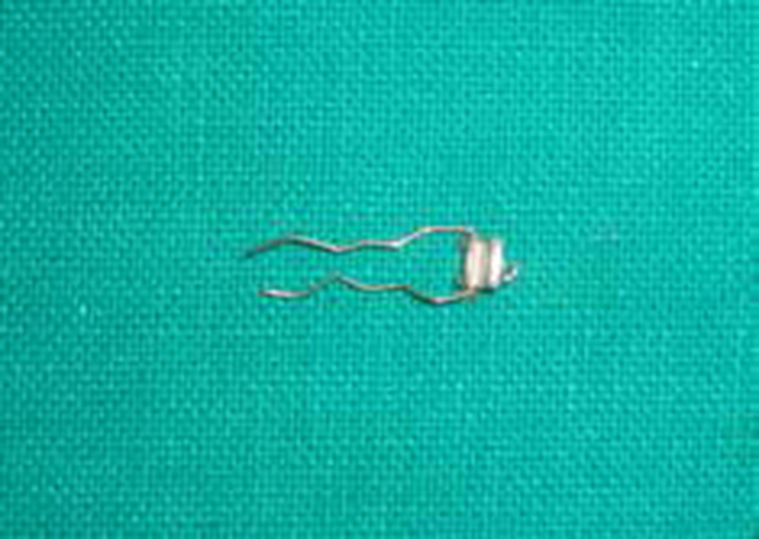
The processed mandibular complete denture was sectioned into two halves at the midline (Figure 5). A hinge assembly was fabricated using an orthodontic bracket with a buccal tube and a 0.7 mm stainless steel wire (Figure 6). Retentive bends were given in the wire for mechanical retention to the denture. Both ends of the wire were incorporated in the right section of the denture on the lingual flange while the bracket was incorporated in the left section at the midline with autopolymerized acrylic resin (Figure 7). Thus, the wire passing through the slot of the buccal tube at the midline facilitated the hinge movement of the mandibular denture inwards (Figure 8).


The patient was instructed about the insertion and removal of dentures and recalled for follow-up visits after 24 hours, 1 week and every 3 months for 1 year. The patient's adaptability was satisfactory with the hinged mandibular denture.
Discussion
The hinge assembly was incorporated on the lingual flange of the mandibular denture so that the denture remained locked at the spread-out position on the residual alveolar ridge without affecting the retention and stability of the denture. An orthodontic bracket, molar tube and stainless steel wire were readily available and easy to maintain in the oral cavity. The only disadvantages would be the bulkiness in the lingual region of the mandibular denture and adequate residual ridge height required in the mid-anterior region to provide space for the attachment of the hinge assembly.
Summary
This design simplified the laboratory technique and reduced the overall cost. It may be useful for patients with reduced mouth opening.