Pazianas M, Miller P, Blumentals WA, Bernal M, Kothawala P A review of the literature on osteonecrosis of the jaw in patients with osteoporosis treated with oral bisphosphonates: prevalence, risk factors, and clinical characteristics. Clin Ther. 2007; 29:1548-1558
Weldon D The effects of corticosteroids on bone: osteonecrosis (avascular necrosis of the bone). Ann Allergy Asthma Immunol. 2009; 103:91-97
FGDP UKUK: The Royal College of Surgeons of England; 2012
Allen MR, Burr DB The pathogenesis of bisphosphonate-related osteonecrosis of the jaw: so many hypotheses, so few data. J Oral Maxillofac Surg. 2009; 67:61-70
Patel V, Kelleher M, Sproat C, Kwok J, McGurk M New cancer therapies and jaw necrosis. Br Dent J. 2015; 219:(5)203-207
Nabil S, Samman N Risk factors for osteoradionecrosis after head and neck radiation: a systematic review. Oral Surg Oral Med Oral Pathol Oral Radiol. 2012; 113:54-69
Assouline-Dayan Y, Chang C, Greenspan A, Shoenfeld Y, Gershwin ME Pathogenesis and natural history of osteonecrosis. Semin Arthritis Rheum. 2002; 32:94-124
Khan AA, Morrison A, Hanley DA Diagnosis and management of osteonecrosis of the jaw: a systematic review and international consensus. J Bone Miner Res. 2015; 30:3-23
Kalsi JS, Abdel-Karim A, Brooke AE A rare case of osteonecrosis in the premaxilla following meningococcal-induced disseminated intravascular coagulation. J Oral Maxillofac Surg. 2012; 70:(12)2814-2818
Jones JP Coagulopathies in the pathogenesis of osteonecrosis. Orthopaeds Trauma. 1997; 11:157-163
Al-Mukhtar Y, Aga F, Hardee P Bisphosphonate related osteonecrosis of the jaw (BRONJ) secondary to an oral ulcer caused by Nicorandil. Br J Oral Maxillofac Surg. 2013; 51:e123-e124
Faibis S, Widmer R, Sapir S, Peretz B, Shapira J Meningococcal septicaemia and dental complications: a literature review and two case reports. Int J Paediatr Dent. 2005; 15:213-219
Khullar SM, Tvedt D, Chapman K, Herlofson BB Sixty cases of extreme osteonecrosis and osteomyelitis of the mandible and maxilla in a West African population. Int J Oral Maxillofac Surg. 2012; 41:(8)978-985
Seamon J, Keller T, Saleh J, Cui Q The pathogenesis of nontraumatic osteonecrosis. Arthritis. 2012; 2012
In the past decade there has been a rise of osteonecrosis of the jaws (ONJ) predominately related to bisphosphonate therapy and osteoradionecrosis secondary to radiotherapy. However, osteonecrosis can occur in the absence of these agents. A case of idiopathic osteonecrosis of the maxilla is reported and the literature discussed.
CPD/Clinical Relevance:It is not always possible to make a definitive diagnosis.
Article
Osteonecrosis of the jaw (ONJ) is identified as ‘non-healing exposed necrotic bone in the maxillofacial region’.1 ONJ has received much publicity in the dental literature recently owing to its induction via various identified medications, eg bisphosphonates, denosumab and bevacizumab, and has now led to the new accepted acronym of MRONJ (Medication Related OsteoNecrosis of the Jaw). Radiotherapy is also a common cause for ONJ and is referred to as OsteoRadioNecrosis (ORN). There remain other causes for ONJ but these are uncommon. A spontaneous case of ONJ due to vascular ischaemia is presented.
Case report
A 94-year-old-male was referred by his general dental practitioner to the Head and Neck clinic at Guy's Hospital due to exfoliation of his upper central incisors and exposed non-healing bone in the premaxilla, as a malignant process was suspected. Prior to exfoliation of the teeth, the patient experienced spontaneous intra-oral bleeding from the maxillary midline region.
A medical history revealed a patient with modest memory impairment. In the past he had undergone a quadruple coronary artery bypass and currently suffered from hypertension. His medication included; bisoprolol, atorvastatin, clopidogrel, aspirin, nicorandil, isosorbide mononitrate, GTN spray, omeprazole, Adcal-D3. He did not smoke or consume alcohol.
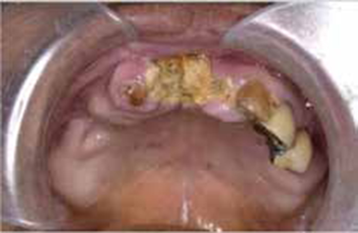
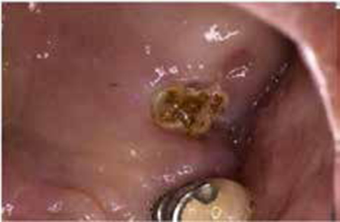
Extra-oral examination was unremarkable. Intra-oral exam revealed two separate areas of exposed necrotic alveolar bone. The largest was in the upper incisor region (Figure 1) with a smaller necrotic area in the upper left first molar region (Figure 2). There was no purulent discharge and the tooth sockets in the pre-maxillary region were intact with no evidence of bony erosion. There was no oro-nasal communication and both oral and denture hygiene were deemed satisfactory.
Figure 1. Presenting image of osteonecrosis upper incisor region.Figure 2. Presenting image of osteonecrosis upper left first molar region.
The appearance was identical to MRONJ or ORN, but these therapies had not been provided and were confirmed by the general medical practitioner. Investigations included; blood assays (full blood count, urine, electrolytes, liver function tests, bone profile, thyroid and parathyroid function tests, auto-antibody screens, ESR, CRP, PSA and sickle cell). No cause for ONJ could be identified.
Imaging (dental panoramic tomograph and a cone beam CT) showed bony destruction of the anterior maxilla back to the molar teeth with right-sided sinusitis. A chest X-ray excluded metastatic disease and tuberculosis and a nuclear medical scan revealed increased activity in the pre-maxilla in line with the confirmed ONJ.
Biopsy of the exposed bone and adjacent soft tissue from the margin of the necrotic area concluded sequestration and non-specific inflammation with no evidence of malignancy. There were no signs of local or systemic infection to indicate osteomyelitis.
A diagnosis of ischaemic osteonecrosis of the pre-maxilla of unknown cause was made by exclusion.
Owing to the patient's deteriorating mental state and the lack of symptoms, a conservative approach was adopted. Under local anaesthetic, the sharp and protruding edges of the exposed bone were reduced using piezoelectric surgery, without the need to raise soft tissue flaps, to avoid diminishing the blood supply further. Debridement was followed by a post-operative course of oral doxycycline (100 mg) and chlorhexidine gluconate 0.2% mouthwash.
On 3-month review, the patient reported no symptoms and examination revealed an improved appearance of the soft tissue with early suggestion of the necrotic bone improving.
Discussion
The pathophysiology of ONJ is complex and poorly understood. Theories include progressive osteocyte destruction resulting from bone and bone marrow infarction secondary to compromised bone vasculature.2 It is this reason which is thought to explain why ONJ is twice as likely to occur in the mandible compared to the more vascularized maxilla.3 Other hypotheses emphasize the role of infections, altered fat metabolism and toxic damage of the soft tissue, amongst others.4
The two most common predisposing factors for ONJ are medication-related or radiotherapy. Oncology patients treated with intravenous bisphosphonates have a higher prevalence of ONJ following tooth extraction, ranging from 1.6–14.8% compared to that of patients prescribed oral bisphosphonates (between 1 in 10,000 to 1 in 100,000).3 Advancements in drug therapy have added further causative agents such as denosumanb and bevacizumab and it is likely that more drugs producing a similar complication are to follow.5 Following radiotherapy the incidence of ORN has ranged between 4.74–37.5%.6 It has been suggested that modern delivery systems with higher precision radiation doses, such as Intensity-Modulated-Radiotherapy (IMR), may reduce the risk of necrosis but there is little evidence to support this supposition at present.
Apart from oncological therapy, sporadic ONJ has been linked with a wide range of factors (Table 1).
Cases describing nicorandil ulceration associated with osteonecrosis have been reported but this was with a background of bisphosphonate use.11 In the current case there was no history of bisphosphonate use and no mucosal ulceration noted prior to this.
In the present case, no explanation could be found except an idiopathic vascular ischaemic event. What is peculiar is that usually the maxilla has a richer blood supply in comparison to the mandible and so is protected from ischaemic insults. The patient was elderly but otherwise well with no underlying pathology that might predispose to infarction of small blood vessels.12 A similar clinical picture has been reported in sub-Saharan Africa.13 Recent studies of the exfoliated specimen suggest an atypical form of tuberculosis (TB) can produce the same clinical picture. The necrotic tissue removed in the present case was reported as negative for TB.
Conclusion
Once iatrogenic events related to cancer therapy are excluded, the rare and sporadic causes of ONJ are mainly related to underlying haematological pathology, fat emboli or haemaglobinopathies leading to clots and subsequent ischaemia.14