White D, Pitts N, Steele J, Sadler K, Chadwick B. Disease and Related Disorders – A Report from the Adult Dental Health Survey 2009.London: The Health and Social Care Information Centre; 2011
2009 ADHS.London: The Health and Social Care Information Centre; 2011
White DA, Tsakos G, Pitts NB, Fuller E, Douglas GV, Murray JJ. Adult Dental Health Survey 2009: common oral health conditions and their impact on the population. Br Dent J. 2012; 213:567-572
Smith BG, Bartlett DW, Robb ND. The prevalence, etiology and management of tooth wear in the United Kingdom. J Prosthet Dent. 1997; 78:367-372
Al-Omiri MK, Lamey PJ, Clifford T. Impact of tooth wear on daily living. Int J Prosthodont. 2006; 19:601-605
Wazani BE, Dodd MN, Milosevic A. The signs and symptoms of tooth wear in a referred group of patients. Br Dent J. 2012; 213
Ahmed KE, Murray CA, Whitters CJ. A prospective survey of secondary care tooth wear referrals: demographics, reasons for concern and referral outcomes. Br Dent J. 2014; 216
Lobbezoo F, Ahlberg J, Glaros AG, Kato T, Koyano K, Lavigne GJ. Bruxism defined and graded: an international consensus. J Oral Rehabil. 2013; 40:2-4
Lavigne GL, Lobbezoo F, Rompré PH, Nielsen TA, Montplaisir J. Cigarette smoking as a risk factor or an exacerbating factor for restless legs syndrome and sleep bruxism. Sleep. 1997; 20:290-293
See SJ, Tan EK. Severe amphethamine-induced bruxism: treatment with botulinum toxin. Acta Neurol Scand. 2003; 107:161-163
Wise M. Citalopram-induced bruxism. Br J Psychiatry. 2001; 178
Lavigne GJ, Rompre PH, Montplaisir JY. Sleep bruxism: validity of clinical research diagnostic criteria in a controlled polysomnographic study. J Dent Res. 1996; 75:546-552
Okeson JP, Phillips BA, Berry DT, Baldwin RM. Nocturnal bruxing events: a report of normative data and cardiovascular response. J Oral Rehabil. 1994; 21:623-630
Gallo LM, Gross SS, Palla S. Nocturnal masseter EMG activity of healthy subjects in a natural environment. J Dent Res. 1999; 78:1436-1444
Kato T, Montplaisir JY, Guitard F, Lavigne GJ. Evidence that experimentally induced sleep bruxism is a consequence of transient arousal. J Dent Res. 2003; 82:284-288
Manfredini D, Ahlberg J, Winocur E, Lobbezoo F. Management of sleep bruxism in adults: a qualitative systematic literature review. J Oral Rehabil. 2015; 42:862-874
Needham R, Davies SJ. Use of the Grindcare® device in the management of nocturnal bruxism: a pilot study. Br Dent J. 2013; 215
Ahmed KE, Murbay S. Survival rates of anterior composites in managing tooth wear: systematic review. J Oral Rehabil. 2016; 43:145-153
Al-Khayatt AS, Ray-Chaudhuri A, Poyser NJ, Briggs PFA, Porter RWJ, Kelleher MG, Eliyas S. Direct composite restorations for the worn mandibular anterior dentition: a 7-year follow-up of a prospective randomised controlled split-mouth clinical trial. J Oral Rehabil. 2013; 40:389-401
Gulamali AB, Hemmings KW, Tredwin CJ, Petrie A. Survival analysis of composite Dahl restorations provided to manage localised anterior tooth wear (ten year follow-up). Br Dent J. 2011; 211
Hemmings KW, Darbar UR, Vaughan S. Tooth wear treated with direct composite restorations at an increased vertical dimension: results at 30 months. J Prosthet Dent. 2000; 83:287-293
Patel M, Seymour D, Chan MF. Contemporary management of generalized erosive tooth surface loss. Dent Update. 2013; 40:222-229
Redman CD, Hemmings KW, Good JA. The survival and clinical performance of resin-based composite restorations used to treat localised anterior tooth wear. Br Dent J. 2003; 194:566-572
Robinson S, Nixon PJ, Gahan MJ, Chan MF. Techniques for restoring worn anterior teeth with direct composite resin. Dent Update. 2008; 35:551-558
Poyser NJ, Porter RW, Briggs PF, Chana HS, Kelleher MG. The Dahl Concept: past, present and future. Br Dent J. 2005; 198:669-676
Burke FJ. Information for patients undergoing treatment for toothwear with resin composite restorations placed at an increased occlusal vertical dimension. Dent Update. 2014; 41:28-38
Milosevic A, Burnside G. The survival of direct composite restorations in the management of severe tooth wear including attrition and erosion: a prospective 8-year study. J Dent. 2016; 44:13-19
al-Quran FA, Lyons MF. The immediate effect of hard and soft splints on the EMG activity of the masseter and temporalis muscles. J Oral Rehabil. 1999; 26:559-563
Okeson JP. The effects of hard and soft occlusal splints on nocturnal bruxism. J Am Dent Assoc. 1987; 114:788-791
Matsumoto H, Tsukiyama Y, Kuwatsuru R, Koyano K. The effect of intermittent use of occlusal splint devices on sleep bruxism: a 4-week observation with a portable electromyographic recording device. J Oral Rehabil. 2015; 42:251-258
Capp NJ. Occlusion and splint therapy. Br Dent J. 1999; 186:217-222
Tooth wear is an increasing problem for general dental practitioners. Attrition is associated with bruxism, primarily a stress-related condition that is difficult to manage dentally. Direct composite restorations are frequently used to restore the worn anterior dentition. Soft occlusal appliances (night guards) are often prescribed in bruxism, despite debatable clinical benefit. Bilaminar (dual-laminate) splints or night guards are composed of two distinct layers of ethylene-vinyl acetate; a soft inner and a harder outer layer. These occlusal appliances are cost-effective to construct, easy to fit and offer greater resistance to occlusal forces than entirely soft occlusal appliances. Patient compliance is excellent. Bilaminar night guards are proposed as an alternative occlusal appliance to prevent further attritional tooth wear from bruxism when TMD is absent and for protection of composite placed to restore the worn dentition.
CPD/Clinical Relevance: The soft occlusal guard is widely used in dentistry but lacks durability and cannot be adjusted. To prevent further attrition and protect restorations, the use of a bilaminar or dual laminate material is advocated in cases of bruxism.
Article
Tooth wear is an increasingly common problem. The Adult Dental Health Survey (2009) reported an increase of anterior tooth wear from 66% in 1998 to 76% of all examined adults in 2009.1 Moderate wear increased from 11% to 15% over the same period. Of particular concern is the reported rise within younger age groups; with 16–24 year-olds displaying a 3% increase in moderate wear to 4% since 1998.2 This trend is likely to lead to more significant management problems in the future.3
Tooth wear can result from multiple processes. A diagnosis of attrition, erosion or abrasion in isolation should only be considered when there is significant clinical or historical evidence to substantiate the diagnosis.4 In reality, tooth wear is often a combination of these diagnoses.5 Attrition is closely related to bruxism, which is widely regarded as a stress-related parafunctional activity. Bruxism and attrition remain a source of great concern for many patients. The principal complaint and driver to seek treatment in 59% of 290 patients referred to a UK teaching hospital for tooth wear was poor aesthetics, followed by sensitivity (40%), functional problems (17%) and pain (14%).6 A similar study reinforced these findings, with aesthetics the primary concern for 54% of patients.7
This paper describes the use of a novel material used as a bilaminar guard to prevent further attrition and protect newly restored teeth in cases of bruxism.
Bruxism
Recently, bruxism has been redefined as repetitive jaw-muscle activity characterized by clenching or grinding of the teeth and/or by bracing or thrusting of the mandible.8 Bruxism occurs during sleep, but daytime (awake/diurnal) clenching is also recognized. The prevalence of bruxism in the population ranges from 5% to 48% and varies according to occupation, smoking habits, alcohol intake, medication and drug addiction.9,10,11 Diagnosing bruxism can be challenging. Whilst patient questionnaires and clinical examinations are important, polysomnography remains the most accurate method for diagnosis.12 This technique is costly, time-consuming and relies upon more advanced instrumentation. Recent literature advises the designation of bruxism into ‘possible’, ‘probable’ and ‘definite’ based on the level of evidence attained with ‘probable’ bruxism relying upon patient recall, questionnaires and clinical examinations.8 Well designed questionnaires can provide significant information and are easy to administer. Study models and clinical photographs may be used to quantify or monitor tooth wear.
Attrition, tooth hypermobility, masticatory muscle hypertrophy, temporo-mandibular disorders (TMD) and fractured cusps may be some of the dental manifestations of bruxism that may assist with diagnosis. Whilst management of these manifestations may be dental, treatment for bruxism is difficult because stress, one of the common causes of bruxism, is a medical problem. It is important to appreciate that stress is generally episodic in nature and so too is bruxism. Temporomandibular joint problems, such as joint sounds (typically clicks), limitation of jaw opening and pain are not necessarily present in patients with tooth wear or in bruxists.
The association between sleep bruxism and sleep pattern is particularly relevant as bruxism was reported to occur during light sleep and was preceded by changes in pulse, breathing and cortical activity.13 Rhythmic masticatory muscle activity (RMMA) of both masseter and temporalis muscles has been monitored using electro-myography (EMG) with simultaneous measurement of cortical and cardiac function by EEG and ECG.14,15 Bruxist episodes can be short bursts of up to 2 seconds of masseteric activity (phasic or grinding) or tonic (clenching) contractions lasting over 2 seconds or a combination of both. Patients should, therefore, be asked about their sleep pattern as this will suggest to the patient a link between sleep, bruxism and tooth wear. Moreover, the association with stress, coping strategies for stress and the limitations of dental management can be discussed.
Numerous general management strategies for bruxism exist.16 Traditional dental management may take the form of soft or hard intra-oral appliances, whilst medical management can include pharmacological agents and cognitive behavioural therapy (CBT). Occlusal appliances (OA), or occlusal splints, may not be the most appropriate management strategy and alternative devices, such as the mandibular advancement appliance (MAA) and a biofeedback device such as Grind-care® could be considered. This biofeedback device uses low-voltage electrical impulses to induce muscle relaxation. Whilst positive results have been published in a small sample, greater research is required.17
Prevention and management of tooth wear
When tooth wear is recorded, identification of the aetiological factor(s) and informing the patient accordingly is fundamental in preventing further wear. It is often easier to manage erosion than attrition as sources of extrinsic or intrinsic acid can be identified, whilst attrition linked with sleep bruxism can pose greater challenges due to its subconscious nature and association with stress. It is important that general dental practitioners highlight the link between stress, bruxism and tooth wear and discuss general coping strategies such as yoga, meditation and exercise. Patient information leaflets can be provided to supplement these discussions.
Multiple studies have discussed and described the use of adhesive restorative materials, such as composite resin, to restore function and aesthetics and help prevent further tooth wear.18,19,20,21,22,23,24 These studies conclude that direct composite restorations are an appropriate medium-term management strategy for anterior tooth wear. The Dahl concept is often utilized with this approach25 and a recent systematic review by Ahmed and Murbay18 supported this approach, with 91% of patients re-establishing posterior occlusion within 18 months following anterior composite restorations at an increased occlusal vertical dimension. Burke devised a patient information leaflet highlighting several important aspects that must be raised with patients prior to undertaking anterior restorations at an increased vertical dimension.26
Several associated factors have been reported in relation to tooth wear and the survival of anterior composite restorations. These include incisor relationship, aetiological cause and lack of posterior support (LOPS); the latter showed a strong correlation (p = 0.003) with failure of anterior composite restorations.27 Failure was also more likely in patients where attrition was the primary aetiological factor and mandibular teeth displayed a higher failure rate over maxillary teeth, potentially due to reduced surface area for bonding. Whilst neither of these findings were statistically significant, they highlight the difficulties faced when managing the worn dentition in bruxists.
Occlusal appliance (night guard) therapy
Patients diagnosed with sleep bruxism, awake bruxism or attrition are often treated with occlusal appliances or night guards to prevent tooth wear and occlusal overload. Various materials and designs exist, with soft silicone/vinyl-based occlusal splints favoured in general dental practice owing to their low cost and ease of construction. Soft splints should be viewed as ‘emergency’ appliances because they are very compressible and may exacerbate chewing/bruxing in severe bruxists.28 Hard occlusal stabilization splints, made from acrylic, are often used in the management of TMD and are not the focus of this article. Hard acrylic splints are often time consuming to produce and fit as they require impressions of both arches, a RCP record, a facebow record and a semi-adjustable articulator. Wax-up of the splints followed by flask investing and heat curing is a time consuming laboratory process, not without potential processing errors and possible chairside difficulty fitting the splint. As discussed above, the traditional alternative is to fit a soft, vinyl-type splint, which only requires one impression. It is compressible and thus easily squeezed during parafunctional activity. Literature exists to show increased masseteric activity with soft occlusal appliance use,29,30 which may be counterproductive in bruxists or those with recent bonded restorations. Furthermore, bruxists can wear through soft splints within a few months. The bilaminar guard has potential benefits over both appliance types discussed above.
Bilaminar (dual-laminate) night guard
The bilaminar (or dual-laminate) splint, more appropriately termed a guard, is an alternative occlusal appliance proposed for the management of attrition-based tooth wear, and protection of anterior composite restorations placed for tooth wear management. Tooth wear in these situations may result from sleep bruxism, awake clenching or a combination of both. These guards are intended for use during the parafunctional episode identified and evidence exists to support the intermittent use of occlusal appliances, which should be considered in patients with bruxism where bruxism may be episodic.31
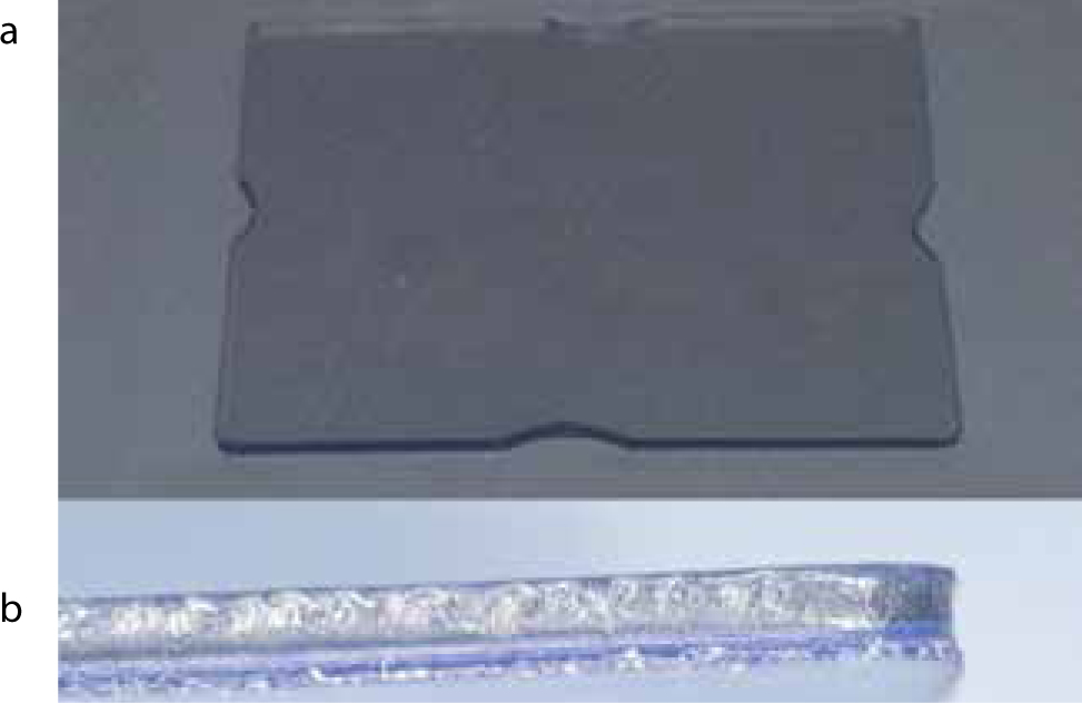
Bilaminar guards are composed of two individual thermoplastic layers, or laminates, chemically bonded to produce a two-layered guard. This results in a soft inner layer often composed of ethylene-vinyl acetate (EVA), and a rigid outer layer composed of hard EVA or polycarbonate (Figure 1). They are vacuum-formed from blanks and are available in a range of different thicknesses (2 mm, 3 mm, 5 mm – Keystone Industries (US)/Metrodent.com(UK), Huddersfield, West Yorkshire/Ortho-Care (UK) Ltd, Saltaire, West Yorkshire), with 3 mm blanks favoured by the authors.
Figure 1.
(a) Flat dual-laminate blank available from Metrodent. com and Ortho-Care UK Ltd in 2 mm, 3 mm and 5 mm. (b) Dual-laminate blanks showing bilaminar design with a soft silicone layer and a hard acrylic layer (5 mm).
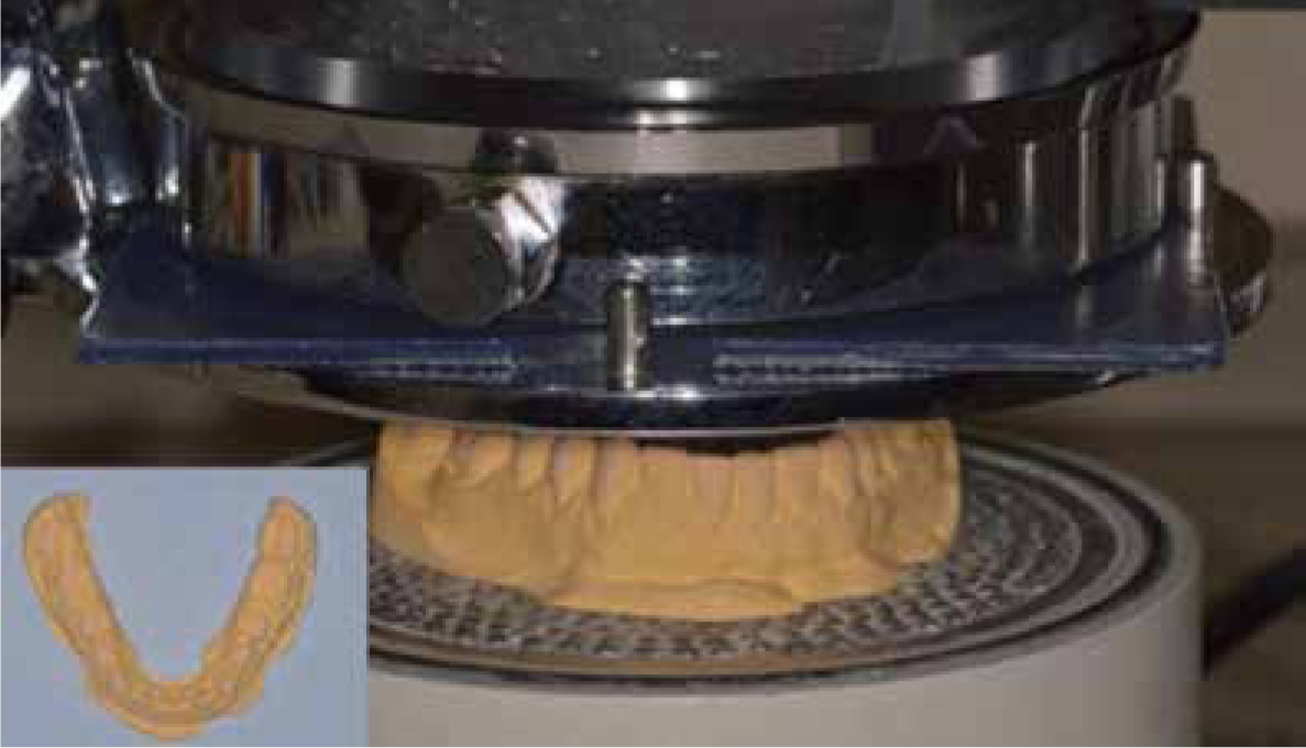
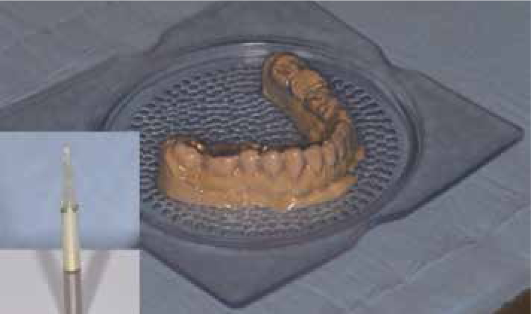
Clinical management begins with an impression of the dental arch planned for guard therapy. Anecdotally, the authors have found mandibular guards easier to construct, with higher patient satisfaction and compliance than maxillary appliances. The cast is modified to remove excess dental stone (lingual/palatal), which would otherwise hinder vacuum-forming (Figure 2). Laboratory vacuum-forming adapts the vinyl to the cast. The resultant guard requires removal from the impression cast with a cutting tool such as a tri-cutter (Figure 3). The laboratory stages are critical, as both temperature and time in the vacuum former must be carefully monitored. Too high a temperature and the material may burn or soften excessively resulting in the guard being too thin and the compressible inner layer being hard. Table 1 summarizes the various laboratory stages.
Laboratory Stages of Production
1
Cast and duplicate impression of dental arch (preferably lower) planned for guard.
2
Remove excess stone from cast to facilitate vacuum–forming. Horseshoe design recommended.
3
Preheat heating element to red hot.
4
Heat dual-laminate blank – soft side first. Avoid over-heating or burning blank.Specific values for temperature and time cannot be given as heating elements vary and thus some degree of trial and error is required. Average heating time is approximately 1–2 minutes.
5
Lower blank onto model and commence vacuum for approximately 2 minutes.
6
Trim with tri-cutter or cutting device.
7
Remove from model and smooth edges.
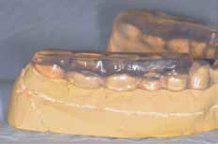
Figure 2. Modified mandibular cast within vacuum-forming machine. Note: heated blank within clamp above model prior to lowering over model. Insert: the modified mandibular cast impression prior to vacuum-forming.Figure 3. A mandibular bilaminar night guard after vacuum-forming has occurred. Insert: tri-cutter bur in straight handpiece used to remove the vacuum-formed splint from the cast model.
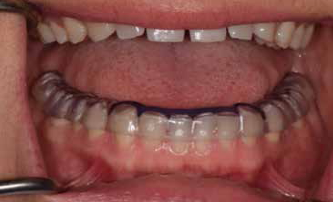
Full occlusal and lingual coverage with half-buccal and labial coverage is the design favoured by the authors to facilitate night guard compliance, retention into the embrasures and optimize gingival health. An in situ mandibular bilaminar splint is seen in Figure 4. The resultant splint is durable, thus providing a greater protective function to prevent further tooth wear and loss of bonded restorations. If required, modification of the occlusal surface can provide thicker acrylic with a flat occlusal plane for those identified as having severe bruxism (Figure 5). This approach requires study casts to be mounted on an articulator. Addition of acrylic requires adjustment at fitting stage to provide a flat occlusal plane with balanced contacts. Splints modified with acrylic can be full– or partial–coverage splints (Figure 5), with the latter not advocated for full-time wear. Complications of partial–coverage splints and uncontrolled tooth movement are discussed elsewhere.32
Figure 4. Mandibular bilaminar splint in situ. Note the early incisal edge and canine tip wear.Figure 5. Bilaminar splint modified with heat-cured acrylic to withstand increased occlusal loads identified in severe bruxists. Note: this is a partial-coverage splint not designed for full-time wear.
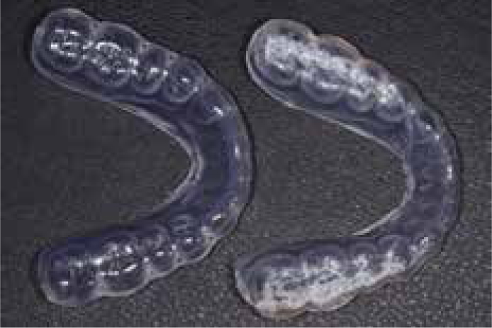
Complications and production errors are rare and similar to soft occlusal appliances. Chairside adjustment is possible with a slow speed acrylic bur in a straight handpiece. Initial hard contacts in inter-cuspal position or retruded contact position are marked with 40 µm thin articulating paper such as Bausch (www.bauschdental.com) and the contacts adjusted extra-orally. Alternatively, the splint can be seated on an articulated study cast mounted in RCP or ICP and adjusted to ensure even occlusal contacts. De-lamination of the splint layers can occur over time. This often occurs at sites where cusp tips have perforated through the hard, outer layer of the night guard following prolonged wear. In these situations, replacement is simple and patients are informed that the guard is protecting the natural teeth and/or restorations by being sacrificial (Figure 6). The rate of wear and damage to the occlusal appliance can be used to assess the severity of bruxism, with rapid delamination or perforation associated with sleep bruxism of greater severity. In these scenarios, alternative management strategies should be considered, eg Biofeedback devices.
Figure 6. On the left is a new replacement bilaminar night guard to replace the year-old cracked and delaminated guard shown on the right. The patient is a 28-year-old female with severe stress-related bruxism of 3 years duration. The previous soft splints lasted on average 3 months. These guards are sacrificed in order to protect the teeth but generally last longer than soft splints.
Discussion
Tooth wear is an increasing problem for the dental practitioner. Alarmingly, greater levels of pathological tooth wear are being reported in younger age groups.1,2,3 Prevention of further wear is fundamental to maintaining optimal health and quality of life. Splint therapy provides one method for distributing the occlusal forces on teeth in those identified with sleep bruxism or daytime clenching. Furthermore, a greater number of bonded anterior composite restorations are being utilized in the management of tooth wear. Survival of anterior restorations is significantly reduced in the presence of a lack of posterior support (LOPS) and, as such, providing posterior support in the form of dentures is beneficial in improving survival of direct adhesive restorations.24 A bilaminar occlusal guard will prevent further tooth wear and/or protect any composite restorations. Guards should be worn during the period of bruxism or clenching. This may be over night (sleep bruxism) or during the day (awake bruxism/clenching), but prolonged wear is not advised.
Soft-silicone/vinyl splints are often used in primary dental care as a management strategy for tooth wear and protection of restorations. It is the authors' opinion that bilaminar splints offer greater wear resistance to occlusal forces than soft appliances; particularly the excessive forces produced during sleep bruxism. Bilaminar guards require minimal clinical and laboratory time, which make them a more viable treatment than hard occlusal stabilization splints. They are also cheap to purchase, construct and replace.
Conclusion
This paper describes the use of a bilaminar intra-oral appliance as a protective guard. Bilaminar guards are proposed as an alternative to soft night guards for use in the protection of anterior bonded restorations and for prevention of further tooth wear in patients diagnosed with attrition and sleep bruxism. They are simple to construct, fit and replace.