
Abstract
Gingival squamous cell carcinomas (SCC) are relatively rare and make up approximately 10% of oral squamous cell carcinomas.
From Volume 44, Issue 10, November 2017 | Pages 984-985
Gingival squamous cell carcinomas (SCC) are relatively rare and make up approximately 10% of oral squamous cell carcinomas.

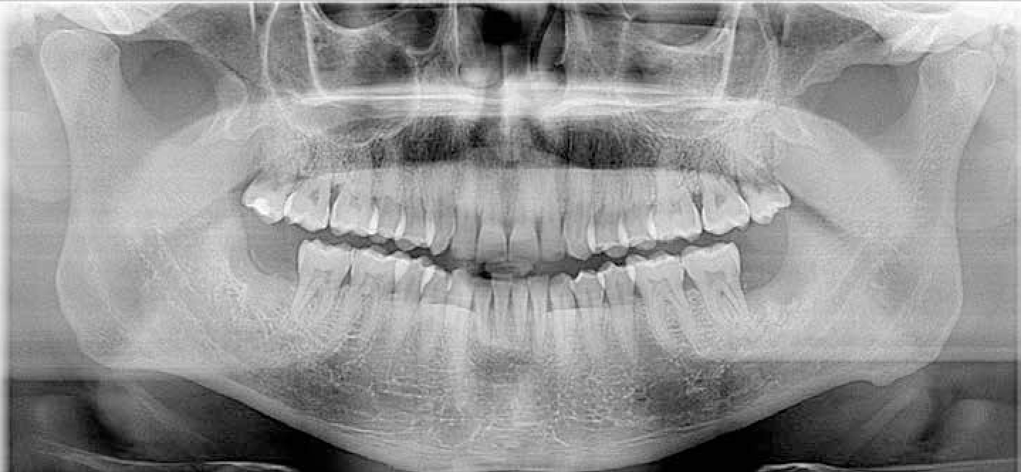
A 39-year-old male patient was referred to the Oral and Maxillofacial Surgery (OMFS) department at Queen Mary's Hospital, Sidcup, by his GDP for the extraction of his lower third molars due to recurrent symptomatic pericoronitis for the past two years. The patient was assessed, examined clinically and radiographically (Figure 1) and booked for routine extractions. His medical history was clear. He smoked around eight cigarettes a day and was an occasional cannabis and alcohol user. His extractions went uneventfully. He had no initial post-operative complications; however, after three weeks he developed left-sided extra-oral facial swelling and was prescribed antibiotics by his general dental practitioner (GDP).
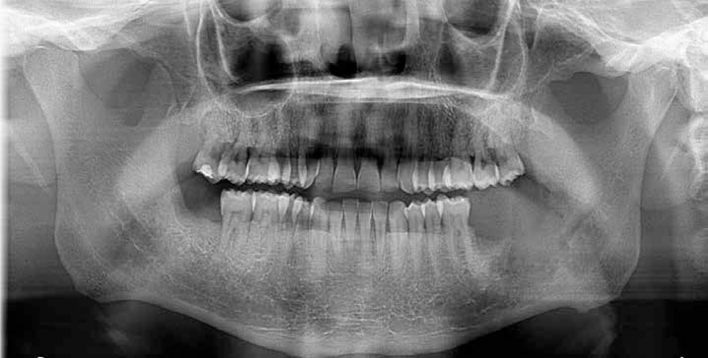
Two months later, he attended the A&E department at King's College Hospital, London, complaining of pain from the lower left quadrant and progressive trismus. He was clinically assessed and examined by the OMFS team and a dental panoramic tomograph (DPT) was taken with no obvious abnormal findings (Figure 2). He was given tongue spatula exercises and was offered a follow-up outpatient appointment, which he declined.
After carrying out tongue spatula exercises for a week with no success, he attended his GDP three weeks later, complaining of trismus. He was diagnosed with masseteric muscular spasm due to bruxism and subsequently fabricated a bite-raising appliance. With his problem persisting, he was seen by his general medical practitioner, who prescribed him diazepam, which alleviated his symptoms temporarily. Six weeks later, he developed another left-sided facial swelling, diagnosed as an infection by his GDP, who then extracted his lower left second molar. He was seen by an osteopath and physiotherapist, whereby the latter carried out laser treatment for his ‘muscular spasm’ and suggested acupuncture. His physiotherapist noticed ‘white material’ from his lower left extraction sockets and suggested he see his GDP, who left him an antibiotic prescription with the receptionist to pick up.
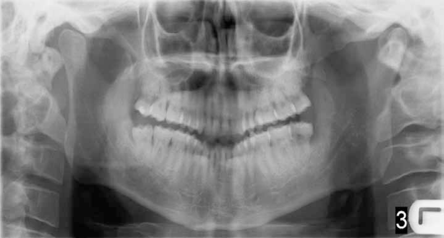
He returned to the A&E department just under two weeks later, with worsening trismus, left lip paraesthesia and exophytic soft tissue growth from the lower left third molar region. A new DPT showed further bony destruction in his lower left quadrant (Figure 3); this too was reflected in a postero-anterior radiograph of his mandible. A CT scan reported ‘clear evidence of osteomyelitis with destruction of the cortex of the angle and body of left mandible.’
He was subsequently admitted as an inpatient at King's College Hospital, and underwent an exploration and multiple biopsies of the left angle of the mandible lesion under general anaesthetic. Antibiotic beads were placed in the extraction socket as a local measure. The biopsies were taken from the exophytic lesion, the mandibular lateral cortex bone and of the periosteal tissue. The specimens were sent to histopathology urgently and reported as poorly-differentiated gingival SCC.
He underwent a left hemimandibulectomy, selective neck dissection and fibula flap with a staging of T4a N1 M0, and was treated by multiple teams including, Head and Neck, Plastic Surgery, Dietitians, Physiotherapy and Speech and Language Therapy. After 12 days he had made a good recovery and was discharged. However, despite commencing a course of radiotherapy and chemotherapy he passed away five months later.
Gingival SCC can be challenging to diagnose which, when delayed, can lead to more invasive treatment and greater morbidity, as seen in this case. In this instance, dentists play a pivotal role in the early detection of gingival SCC. The 5-year survival rate of gingival SCC is significantly less compared to SCC at other sites.2 All healthcare professionals should have increased knowledge and awareness that persistent non-healing oral lesions, including extraction sockets, are to be considered suspicious and managed effectively with appropriate referrals.