Article
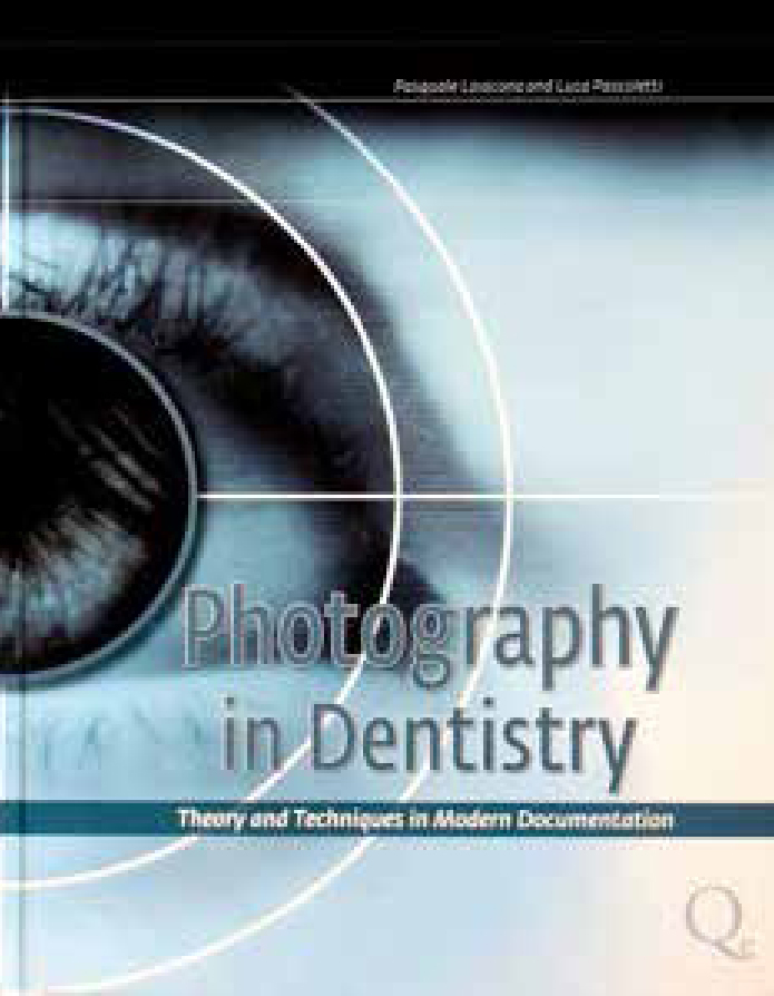
Photography in Dentistry
This is a quality, well produced book consisting of over 300 pages illustrated with over 800 excellent quality images and graphics. I was impressed with the easy layout, which facilitated ‘skipping’ some chapters.
The first chapter covers general principles of photography, with the first few paragraphs enthusing on the indispensible benefits of image capture. There is to my mind a little too much detail for most, but to the enthusiast the detail is interesting. A brief description of aperture and shutter speed in general photography terms follow. This chapter also covers correct handling and choice of camera.
Chapter 2 gives more ‘in depth’ descriptions of optical systems I suspect many will want to skip, that is until the detail on ‘magnifications’, which is good, is reached. The authors have a good knowledge of the main aspects of optical reproduction and the information is accurate. Mastering magnification combined with the camera settings, covered in Chapters 6 and 10, is paramount to achieving consistent reproducible images.
Chapter 3 deals with exposure, and although it includes ISO and, briefly, colour settings, it doesn't cover the association between the main elements of exposure. This comes later in an interesting chapter but which could be skipped.
The first part of Chapter 4 can be skipped, however, later it covers file formats which gives good information on the RAW format and its implementation in clinical photography, and also image transfer for storage.
Chapters 5, 6 and 7 cover the role/use of photography in clinical practice, such as a diagnostic instrument, communication aid and medico-legal use. Camera settings and techniques are also well covered.
Flash units are covered in Chapter 8, with good descriptions of some flash options and the results obtainable. Photography of radiographs is covered in Chapter 9.
The first chapter under the heading ‘Techniques’ is Chapter 10 and covers equipment and accessories, cameras, flash, retractors, etc.
Extra-oral and intra-oral photography techniques are covered in Chapters 11 and 12, giving good practical advice on positioning and camera settings, and good hints and tips for positioning of the operator, camera and patient.
Chapter 13, entitled ‘Documentation’, does a good job of suggesting documentation for orthodontic, periodontal and prosthetic disciplines.
I think most practitioners will want to skip chunks of some of the chapters, but this is easy to navigate. Having said that, the majority of chapters are very useful for the novice and experienced dental photographer alike.

