1. Sore gums

From Volume 41, Issue 5, June 2014 | Pages 471-472

Authors

Crispian Scully

CBE, DSc, DChD, DMed (HC), Dhc(multi), MD, PhD, PhD (HC), FMedSci, MDS, MRCS, BSc, FDS RCS, FDS RCPS, FFD RCSI, FDS RCSEd, FRCPath, FHEA

Bristol Dental Hospital, Lower Maudlin Street, Bristol BS1 2LY, UK

Articles by Crispian Scully

Dimitrios Malamos

DDS, MSc, PhD, DipOM

Oral Medicine Clinic, National Organization for the Provision of Health Services (IKA), Athens, Greece

Articles by Dimitrios Malamos

Article

Professor Crispian Scully
Dr Dimitrios Malamos

A 57-year-old IT negotiator complained of tender gums generally, first noticed about 5 years previously. The condition was persistent, worsening through the day, and sometimes associated with gingival bleeding and a salty taste. The condition was worse in the anterior mandible, and always facially. He denied any blistering or ulcers on the gingivae or elsewhere. Treatment with benzydamine and chlorhexidine mouthwashes and low dose corticosteroid mouthrinses (betametasone) had not significantly improved his symptoms.

There were no cutaneous, gastro-intestinal, genital, ocular or joint problems (apart from traumatic arthritis) and no history of fever. The medical history included knee arthritis from sports. He had a family history of hypercholesterolaemia. There were no other cardiorespiratory or bleeding problems. The patient was on medication with citalopram (for a work-related stress episode) and simvastatin, and had no known allergies. The social history included no tobacco use but alcohol consumption at 60 units weekly. Liver function had been normal when tested twice in the past year.

Extra-oral examination revealed no significant abnormalities and specifically no pyrexia, cervical lymph node enlargement, nor cranial nerve, salivary or temporomandibular joint abnormalities. He had masseteric hyperplasia bilaterally.

Oral examination revealed a dentition which was well preserved. There was clinical evidence of periodontal attachment loss and pocketing. He had extensive desquamation in several areas both facially (Figure 1) and lingually/palatally, with erosions bilaterally, especially posteriorly, and also palatal to the maxillary molars. Plaque control was lacking. Examination of the mucosae showed no other lesions except a crenated tongue margin.
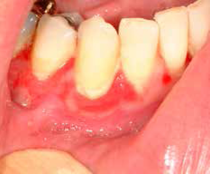
Figure 1. Extensive gingival desquamation.

What is the single most likely diagnosis?

  • Herpetic gingivitis;
  • Erythema multiforme;
  • Lichen planus;
  • Vesiculobullous disease (pemphigoid/pemphigus).
  • The answer to what is the single most likely diagnosis?

    d) Vesiculobullous disease: this can involve the gingivae in middle-age, causing chronic erythema and desquamation. The absence of skin or other mucosal lesions here suggests pemphigoid rather than pemphigus. The history and clinical findings were most consistent with a diagnosis of mucous membrane pemphigoid, since there were, and had been, no other lesions orally or elsewhere. However, it would be prudent to exclude pemphigus by biopsy/immunostaining. The patient had pemphigoid and was treated initially with a betametasone mouthwash at least 3 times daily for at least 3 minutes each time, for one month, with oral hygiene instruction, as plaque aggravated the symptoms. His GDP constructed soft acrylic full coverage splints extending into the sulci, for use of steroid (clobetasol) ointment overnight; this helped suppress the symptoms. An ophthalmological opinion was arranged, since conjunctival lesions may arise in some patients with pemphigoid.

  • Herpetic gingivitis is characterized by an acute diffuse gingival erythema, ulcerations and vesicles and is accompanied by fever and cervical lymphadenopathy. In contrast, the gingivae in our patient are characterized by chronic erythema (>5 years) with extensive desquamation rather than ulcerations without vesicle formation and without general symptomatology.
  • Erythema multiforme is characterized by acute episodes of extensive ulcerations throughout the oral mucosa, often seen in young patients but with no pyrexia, cervical lymphadenopathy and malaise. Our patient had gingival lesions at the age of 52, without free periods from the disease, and his lesions were found strictly in his gums and not in other parts of his mouth skin and other mucosae.
  • Lichen planus is a chronic mucocutaneous disease which rarely affects only the gingivae, showing desquamation alone or in combination with white lesions. In our case no white lesions were noted.
  • Which laboratory investigation(s) is/are mandatory for the diagnosis?

  • Histopathological examination;
  • Immunofluorescence (direct/indirect);
  • Blood tests (haematological, biochemical, immunological);
  • Cell culture.
  • The answer to which laboratory investigation(s) is/are mandatory for the diagnosis?

  • Routine histopathological examination is useful as it determines if the lesion arises from the epithelium or from the underlying connective tissue. The disorders are characterized by the following:
  • Pemphigus by intra-epithelial acantholysis;
  • Pemphigoid by subepithelial bullae;
  • Lichen planus by a dense zone of chronic inflammatory cells at the upper part of corium;
  • Herpetic gingivitis and erythema multiforme show changes within the epithelium and lamina propria (‘interface dermatitis’).
  • Direct immunofluorescence may help distinguish pemphigus from pemphigoid by the deposition of IgG, or IgA between epithelial cells in pemphigus or along the epithelial basement zone in pemphigoid.
  • Haematological tests, such as white blood count, and biochemical tests, such as blood sugar, thyroid hormones, or antibodies for viruses are rarely helpful for the diagnosis.
  • Cultures are useful only for inoculation and growth of bacteria, fungi or viruses from the gingivae but here provide little help for the diagnosis.