Article


A 57-year-old woman was referred for evaluation of a soft polypoid lesion in her upper left alveolar mucosa that had been present for 2 months. There was slight tenderness over the left maxillary sinus, a salty taste and occasionally nasal bloody discharge, but no pain, eye problems or regional lymphadenitis. Her medical history was clear.
On extra-oral examination there was slight tenderness on palpation over the left maxillary sinus.
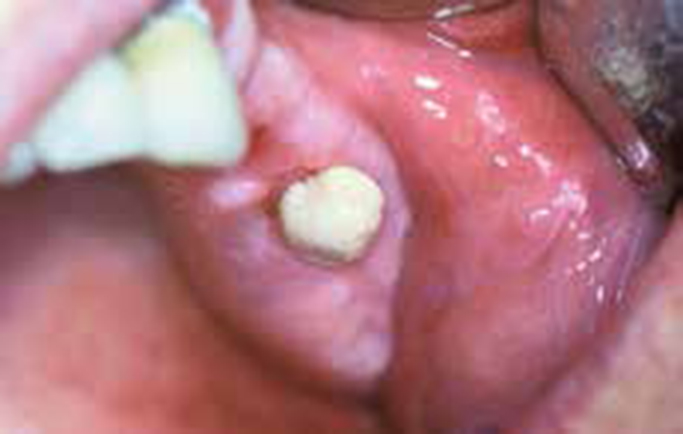
On oral examination, a polypoid, soft, non-tender mass was seen in the area of the left maxillary first molar, which had been extracted 3 months previously (Figure 1). The lesion had a whitish-yellowish colour and was easily lifted from the alveolar mucosa and bled with slight pressure. Radiography showed a hazily radio-opaque maxillary left sinus with a bony discontinuity of its floor at the area of the extracted molar.

Q1. Which is the probable diagnosis?
A1. The answer to which is the probable diagnosis?
(a) Parulis is a soft erythematous lesion that appears on the gingiva in association with a non-vital tooth at the site of the intra-oral drainage of a dental abscess. This lesion has a red colour as it is a granulation tissue. Although pus or blood can exude from the lesion, taste changes and nasal discharge are rare. These findings, together with the absence of a non-vital tooth in this patient, and with the clinical and radiographic characteristics, allow exclusion of a parulis.
(b) Rhinocerebral mycormycosis is a severe infection of the nose and antrum by a Rhizopus fungus, typically seen in patients with uncontrolled diabetes or an immunodeficiency. Two types are recognized, acute and chronic. The chronic type is characterized by a maxillary swelling and causes eye problems, such as ptosis, proptosis and vision loss. Although the lesion in question is chronic, it is not associated with alveolar bone necrosis or eye symptomatology and the patient was otherwise healthy.
(c) Antral osteomyelitis is an infection and inflammation of the maxilla which extends upwards into the antrum. It is characterized by severe pain, swelling, fever, cervical lymphadenopathy, a draining sinus, periosteal thickening and bone destruction with sequestra formation. Maxillary osteomyelitis is rare and the absence of severe pain and swelling or pus and bony sequestration and general symptomatology in this patient excludes this.
(d) Antral tumours are either benign, such as inverted papillomas or ameloblastomas, or malignant, such as carcinomas, adenoid cystic carcinomas, lymphomas and sarcomas and can destroy the maxillary antrum floor and enter the oral cavity. The various tumours (benign or malignant) may, at an early stage, be asymptomatic or cause only epistaxis or nasal obstruction but, as the tumour grows, can cause severe bone destruction, visual problems and cranial neuropathy. In this patient, the lesion had been noticed for only two months and caused mild pain and nasal discharge, but not cranial nerve or visual dysfunction. Radiography did not show bony destruction or a mass in the antrum suggesting a tumour.
(e) Oro-antral fistula is a pathological communication between the oral cavity and the maxillary sinus, which has its origin from iatrogenic complications such as a tooth extraction. This canal is lined either by oral or antral epithelium and filled with immature granulation tissue and presents as a soft polyp in the area of the extracted tooth. The clinical and radiographic characteristics in this patient match those of an oro-antral fistula.
