Article


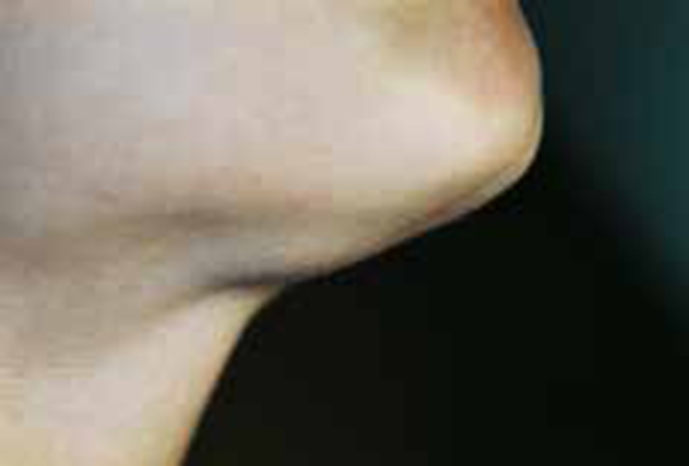
A 9-year-old boy attended complaining of a persistent single swelling in the floor of his mouth causing some difficulties with eating, swallowing and speaking. There was no previous trauma to this area. None of his close relatives had similar lesions. Examination of the skin below the chin revealed a small swelling first noticed by his mother 3 weeks previously (Figure 1). There was no redness, tenderness or sinus tract or cervical lymph node enlargement.

Oral examination revealed a large dome-shaped, non-tender, fluctuant, transparent swelling in the floor of his mouth (Figure 2). This swelling extended bilaterally, beneath the salivary ducts, and pushed the tongue upwards.

Radiography and ultrasonography were undertaken and the lesion, when removed under GA, was found to be composed of granulation tissue lining a cavity containing mucus, muciphages and inflammatory cells. There was no recurrence by two-year follow-up.
Q1. Which is the probable diagnosis?
A1. The answer to which is the probable diagnosis?
(a) Dermoid cyst is a true hamartoma caused when skin and skin structures become trapped during foetal development. The dermoid cyst presenting in the floor of the mouth is rare but represents the second most common after the lateral eyebrow dermoid cyst. There are 3 types: i) dermoid or compound; ii) teratoid and iii) epidermoid cysts, according to the presence or not of skin and skin appendages within the fibrous cystic wall. The patient's lesion has some features in common with the dermoid cyst, such as the location, age and sex predilection, but was different in consistency and histological characteristics. The lesion was more fluctuant and composed of granulation tissue without an epithelial lining, while the dermoid cyst (especially the epidermoid type) has a more rubbery consistency and its cyst wall is fibrous with an epithelial lining. Both lesions show a cystic structure on ultrasonography. The CT scan can differentiate the two lesions using the pathognomonic criterion of a ‘sack of marbles’ appearance seen in dermoid cysts.
(b) Branchial cleft cyst is a congenital lesion arising on the lateral side of the neck, from a failure of obliteration of the second branchial cleft during embryonic development. This cyst is asymptomatic and appears later in life with the characteristic histological features, such as an epithelial lining and the presence of lymphoid tissues in germinal centres. In contrast, the lesion in this case is located on the floor of the mouth and presented at a younger age, but lacked the histological features of a branchial cyst.
(c) Lipoma is a benign tumour of adipose tissue which can be found in the floor of the mouth. As in this lesion, lipoma is an asymptomatic lesion with no sex and race predilection but differs in colour, consistency and histology. This lesion is fluctuant with slight bluish colour and contains mucus rich in amylase and abundant macrophages and inflammatory cells on aspiration. The lipoma has a more elastic structure, yellow colour and contains a plethora of adipocytes and precursor fat cells with increased levels of lipoprotein lipase on aspiration. This lesion is surrounded by a fibrous capsule and composed of fatty cells, features not seen in the current patient.
(d) Lymphangioma is a rare benign hamartomatous lesion of the lymphatic system involving the tongue and floor of the mouth and usually present at birth. This lesion can be superficial and has a pebbly appearance with clear or bluish colour, or can be deep, causing a neck swelling. Superficial lesions consist of dilated lymph vessels lined by endothelial cells close to the oral epithelium. Deeper lesions are composed of irregular lymphatic vessels forming cysts containing eosinophilic proteinaceous fluid. The absence of lymphatic vessels in the current lesion excludes lymphangioma.
(e) Ranula is a large mucocele in the floor of mouth, caused by extravasation of mucus superior to mylohyoid muscles due to trauma or obstruction of the duct of the major salivary glands. It is a painless, non-fixed swelling interfering with swallowing and speaking. It is a cystic lesion whose lumen wall is made only by a granulation tissue without epithelial lining and its fluid contains mucus rich in amylase, macrophages and inflammatory cells. This case has all the clinical and histological characteristics of a ranula.
