Article


A 20-year-old female secretary attended for tooth whitening. Her medical history was clear. Her social history revealed a relatively normal diet with a lot of fruit juices but with no tobacco or alcohol consumption.
Extra-oral examination revealed no significant abnormalities, such as cervical lymph node enlargement, cranial nerve, salivary or temporomandibular joint.
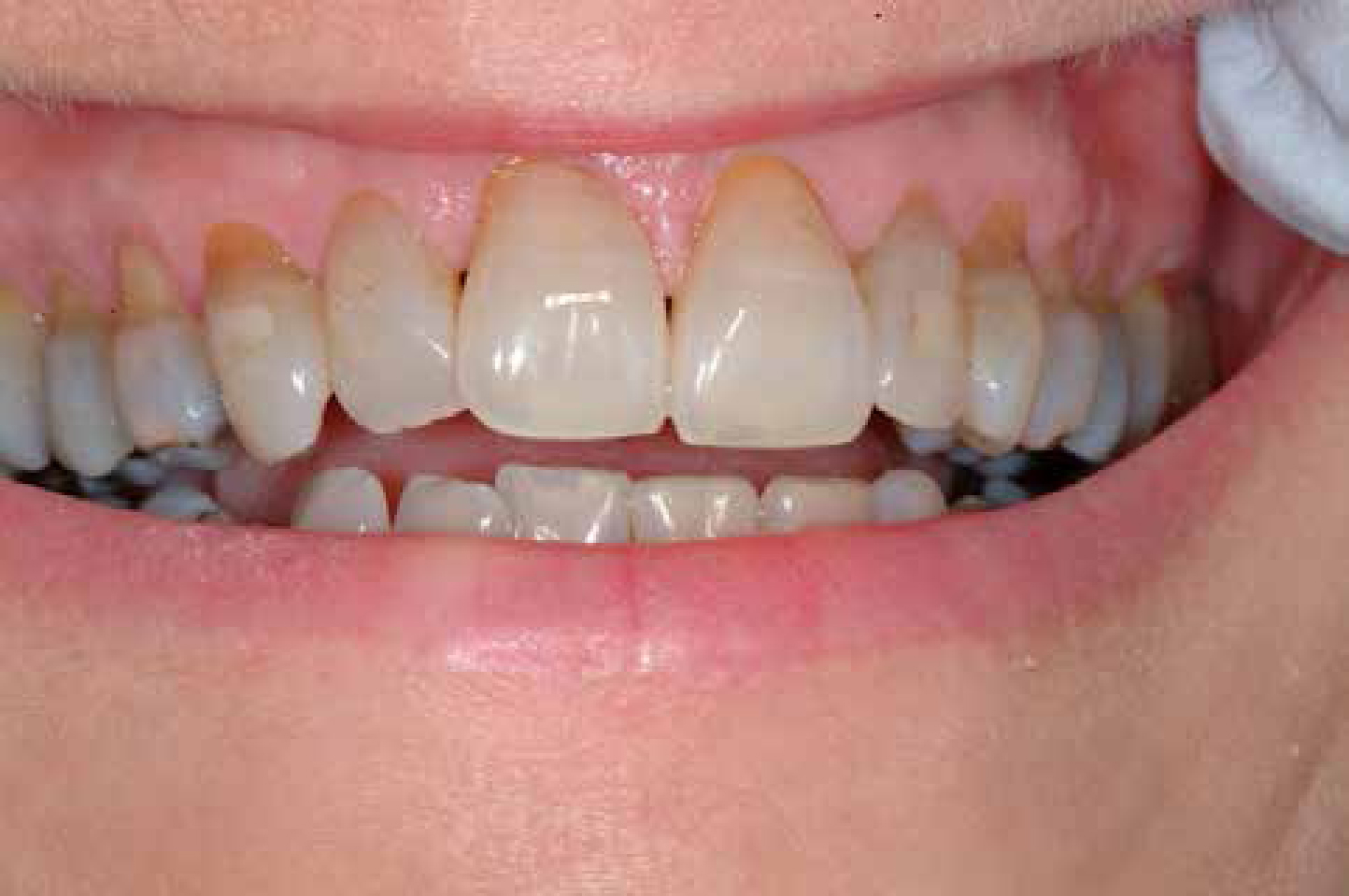
Her dentition was as shown (Figure 1) with tooth discoloration and surface loss, but with no clinical evidence of periodontal attachment loss or pocketing.

Q1. Which is the main cause of such teeth discoloration?
A1. The answer to which is the main cause of such teeth discoloration?
c) Tetracycline staining is the cause here. Tetracycline given in pregnant mothers or in children <8 years old causes a diffuse discoloration in deciduous and permanent teeth, respectively, as it is incorporated into the hard tissues (bones, teeth) that are calcifying at that time. The discoloration begins as bright yellow and, with UV light exposure, finally becomes diffuse brown but without enamel hypoplasia. Our patient had a diffuse brown discoloration of all her permanent teeth, which did not show any change in the shape, texture and mineralization. (a) With ageing, the permanent are darker than the deciduous teeth, as these teeth are exposed over the years to external stains such as those from tobacco, tea or coffee and chlorhexidine mouthwashes. In addition, the permanent teeth often suffer attrition and abrasion, exposing the dentine, which finally increase the tooth discoloration.
(b) Fluorosis causes enamel discoloration resulting from hypomineralization due to excessive ingestion of fluoride (drinking water, mouthwashes, toothpastes) during enamel maturation. This hypomineralization causes chipping of the enamel and yellow to brown discoloration.
(d) Dentinogenesis imperfecta is uncommon and characterized by brown-blue appearance of teeth with enamel which chips off easily and teeth prone to occlusal wear. Osteogenesis imperfecta is due to a deficiency of a type 1 collagen formation, causing fractures of bones, blue sclera and dentinogenesis imperfecta. Our patient had no history of bone fractures or blue sclera.
(e) Erythropoietic porphyria is a congenital disease of haem metabolism causing incorporation of the porphyrin pigments into teeth during dental formation and producing a pink diffuse discoloration of all teeth. Under ultraviolet light the teeth in porphyria stain red but, in contrast, in tetracycline staining they look yellow.
Q2. Which is the best treatment?
A2. The answer to which is the best treatment?
(c) Bleaching in the dental office initially, by application of various whitening agents such as hydrogen peroxide, is usually the preferred treatment. Where bleaching is not indicated, dental bonding can be used to improve the appearance of the teeth by applying composite resin filling on the surface of the teeth under intense cure light. The main problem is the bonding is not permanent and must be followed up.
(a) Removing the external stain from tea, coffee or beverage, calculus and chlorexidine use, by scaling and polishing, results in the brown colour remaining as the stain being inside the teeth.
(b) Enamel microabrasion consists of a rotary application of a mixture of weak hydrochloride acid and silicon carbide particles in a water soluble paste. This is widely used in simple localized fluorosis cases.
(e) Prosthetic restorations as partial (laminate veneers) or full crowns can be used in severe cases with extensive teeth discoloration and in patients very worried about their appearance.
