Article


An 18-year-old female university student was referred by the Student Health Service after the dental officer spotted some sublingual lesions at a routine dental examination. The patient was symptomless with a clear medical history. There were no cardiorespiratory, bleeding, eating, anogenital, gastro-intestinal, eye, skin, or joint problems. No history of fever or known allergies and medication apart from OCP was reported. Her alcohol consumption was from 5 to 50 units weekly but there was no current tobacco use.
Extra-oral examination revealed no significant abnormalities and specifically no pyrexia, cervical lymph node enlargement, or cranial nerve, salivary, masticatory muscle (masseters, temporalis, medial or lateral pterygoids), or temporomandibular joint abnormalities.
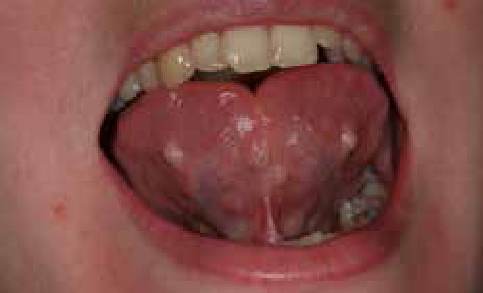
Oral examination revealed a dentition that was basically intact. There was no clinical evidence of periodontal attachment loss or pocketing. Six discrete, painless, white/pink nodules with clear fluid or depressions centrally were noted in the ventral surface of the tongue; four are visible in Figure 1. The right maxillary central incisor was chipped.

Q1. Which is the likely cause of these lesions?
A1. The answer to which is the likely cause of these lesions?
(a) Piercing. This is now quite commonplace but rarely seen in such a number. The placement in a fairly symmetric pattern is a giveaway (Figure 1). Some patients have the habit of inserting foreign material, like pins, into the tongue, which can cause chronic irritation responsible for the development of mucous retention cysts (nodules with clear fluid) or pyogenic granulomas or fibrous keloid-like lesions. The symmetrical distribution of these particular lesions confirms the clinical suspicion that these lesions have been induced by the patient with piercing. This habit was also responsible for the fractures of the incisive edges in our patient.
(b) Virus infection can cause discrete nodules; strains of the human papillomavirus (HPV) family are responsible for papillomas (HPV 6 and 11), warts (HPV 2 and 4), condylomata accuminata (genital warts) (HPV 6 and 11) or focal or multifocal epithelial hyperplasia (HPV 13 and 32). These HPVs are not typically associated with oral or oropharyngeal cancer – caused by HPV 16, HPV 18 or others. Also, the molluscum contagiosum, a DNA poxvirus, is a very rare cause of multiple nodules in the mouth, which look like a dome, smooth surfaced but with dimpled centres affecting the skin, mainly children or HIV-infected people.
(c) Sarcoidosis can cause multiple painless nodules scattered in the mouth, together with a few erythematous nodules of the dorsum of hands and feet and numerous granulomas in the lungs and lymph nodes.
(d) Multiple endocrinopathy syndrome is a rare inherited disease in which one or more of the endocrine glands (pancreas, parathyroid and pituitary) form a tumour. Three types (1, 2A and 2B) are recognized but only the type 2B has oral mucosal lesions (neuromas).
(e) Cowden syndrome is a rare autosomal, dominant disease (PTEN gene) characterized by multiple skin and oral mucosa hamartomas and nodules, thyroid and breast anomalies, together with polyposis of the gastro-intestinal tract.
(f) Multiple mucoceles can be seen but are uncommon.
