Article
We present a curious case of a patient referred by the GDP to the local Oral and Maxillofacial unit. The patient had been urgently referred due to an exophytic growth in a post-operative LR6 extraction socket. Incidentally, he had been complaining of a more posterior discomfort to the ipsilateral side of his mouth. Initial examination showed a mild trismus and a small granulomatous lesion overlying the extraction socket.
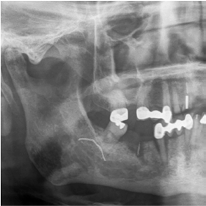
An OPG was taken (Figure 1) that showed two anomalies. The first, within the extraction site, appeared to show a retained foreign body – likely a root-filling material. The second, in the region of the angle of the mandible, appeared densely radio-opaque and coiled or braided.

A biopsy was performed to the LR6 socket tissue and the socket explored and debrided. All tissue obtained was sent for histology, which later confirmed granulation tissue. Careful re-examination of the patient, focused to the right posterior lingual sulcus, revealed a large coiled brush with a central malleable metal process. Initially, it had been missed, as it lay between the palato-glossal arch and the base of the tongue. The object was removed without complication and quickly identified as a large interdental brush, whose handle had separated. The adjacent mucosa was examined and found to be mildly erythematous but with no ulceration. Immediate relief followed the removal of the brush.
We felt it prudent that dental professionals are aware of the potential for work hardening of the malleable metal of interdental brushes. In this case, it ultimately led to a fracture separation of the handle. Patients need to be made aware of the finite span of these brushes when being advised on their use.
