Oral medicine: 12. lumps and swellings: salivary

From Volume 40, Issue 9, November 2013 | Pages 778-779

Authors

David H Felix

BDS, MB ChB, FDS RCS(Eng), FDS RCPS(Glasg), FDS RCS(Ed), FRCPE

Postgraduate Dental Dean, NHS Education for Scotland

Articles by David H Felix

Jane Luker

BDS, PhD, FDS RCS, DDR RCR

Consultant and Senior Lecturer, University Hospitals Bristol NHS Foundation Trust, Bristol

Articles by Jane Luker

Crispian Scully

CBE, DSc, DChD, DMed (HC), Dhc(multi), MD, PhD, PhD (HC), FMedSci, MDS, MRCS, BSc, FDS RCS, FDS RCPS, FFD RCSI, FDS RCSEd, FRCPath, FHEA

Bristol Dental Hospital, Lower Maudlin Street, Bristol BS1 2LY, UK

Articles by Crispian Scully

Article

David H Felix
Jane Luker
Crispian Scully

Specialist referral may be indicated if the Practitioner feels:

  • The diagnosis is unclear;
  • A serious diagnosis is possible;
  • Systemic disease may be present;
  • Unclear as to investigations indicated;
  • Complex investigations unavailable in primary care are indicated;
  • Unclear as to treatment indicated;
  • Treatment is complex;
  • Treatment requires agents not readily available;
  • Unclear as to the prognosis;
  • The patient wishes this.
  • Salivary gland swelling

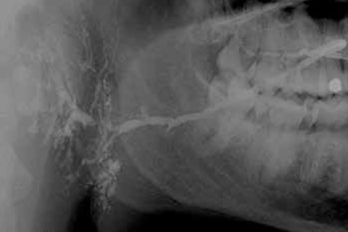
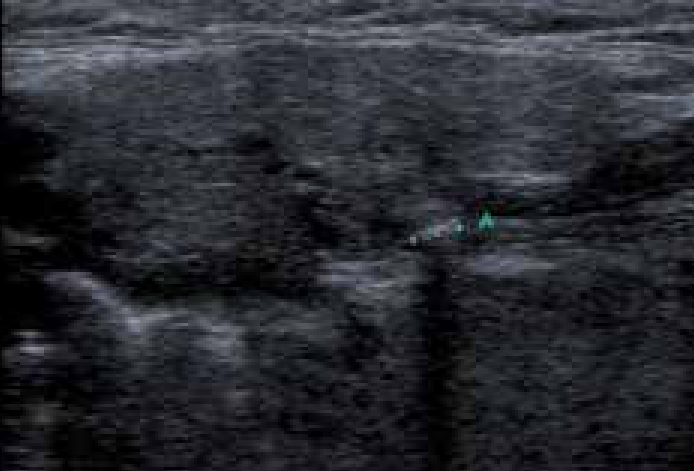
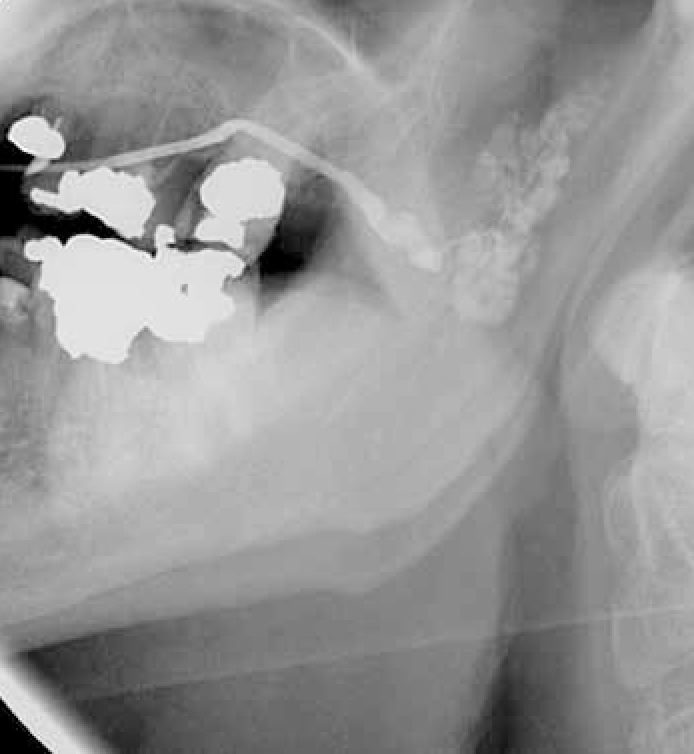
    Salivary glands usually swell because of inflammation (sialadenitis) (Figure 1), which is often viral but may have other causes (Table 1). Obstruction of salivary flow is another common cause (obstructive sialadenitis) (Figures 2, 3). Rare causes include salivary gland or other neoplasms (Figures 47).


  • Inflammatory
  • Mucoceles
  • Mumps
  • Ascending sialadenitis
  • Recurrent parotitis of childhood
  • HIV parotitis
  • Other infections (eg tuberculosis)
  • Sjögren's syndrome
  • Sarcoidosis
  • Cystic fibrosis
  • Neoplasms (mainly pleomorphic salivary adenoma, but also monomorphic adenomas and malignant tumours)
  • Duct obstruction (eg calculus)
  • Sialosis (usually caused by autonomic dysfunction in starvation, bulimia, diabetes, or alcoholic cirrhosis)
  • Deposits rarely (eg amyloidosis and haemochromatosis)
  • Drugs rarely (eg chlorhexidine, methyl dopa, phenylbutazone, iodine compounds, thiouracil, catecholamines, sulfonamides, phenothiazines and protease inhibitors
  • Figure 1. Right parotid sialogram showing dilation of the intraglandular ducts consistent with sialadenitis.
    Figure 2. Ultrasound of submandibular gland, stone identified (A).
    Figure 3. Left parotid sialogram showing intraglandular ductal dilation and a filling defect at the hilum of the gland consistent with a parotid calculus.
    Figure 4. Parotid neoplasm.
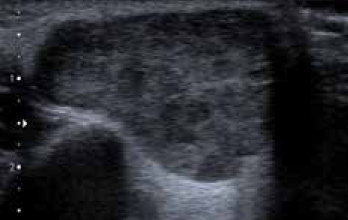
    Figure 5. Well defined area of hypo-echogenicity within salivary tissue consistent with a parotid neoplasm.
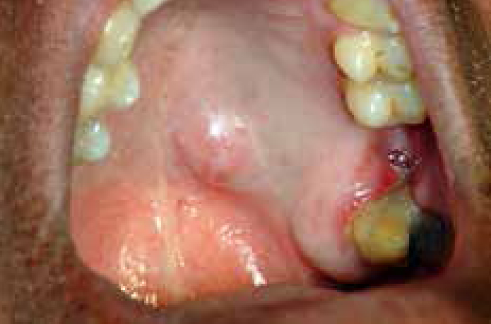
    Figure 6. Minor salivary gland neoplasm.
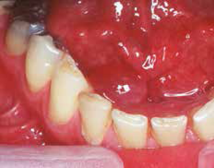
    Figure 7. Malignant salivary gland neoplasm.
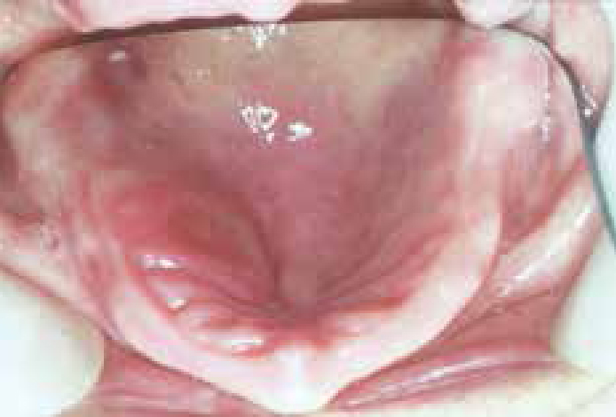
    Figure 8. Mucocele, floor of mouth (ranula).

    In children, most salivary gland swellings are caused by mucoceles or mumps. In adults, most swellings of the salivary glands are caused by mucoceles (Figure 8), salivary duct obstruction (typically by a stone); but sialadenitis, Sjögren's syndrome and neoplasms are important causes to be excluded.

    Diagnosis of salivary gland swelling

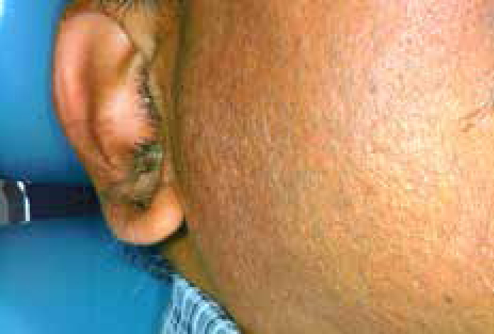
    It can be difficult to establish whether a major salivary gland is genuinely swollen, especially in obese patients. A useful guide to whether the patient is simply obese or has parotid enlargement is to observe the outward deflection of the ear lobe, which is seen in true parotid swelling.

    Diagnosis of the cause is mainly clinical but investigations such as imaging (especially ultrasound), liver function tests, serology for viral antibodies autoantibodies or biopsy, may be indicated.

    Management

    A specialist opinion is usually needed and treatment is of the underlying cause.

    Immediate treatment is needed for acute bacterial sialadenitis; under ideal conditions antimicrobial therapy should be determined by results of culture and sensitivity of a sample of pus from the duct. However, as first line therapy a penicillinase-resistant penicillin such as flucloxacillin is appropriate. In patients with penicillin allergy, erythromycin is a suitable alternative. In addition, general supportive measures such as analgesia and increased fluid intake are important. Thereafter, specialist referral is generally indicated to identify any predisposing factors. Neoplasms need Specialist attention.