Oral medicine: 14. radiolucencies and radio-opacities. a. bone diseases

From Volume 41, Issue 1, January 2014 | Pages 84-87

Authors

David H Felix

BDS, MB ChB, FDS RCS(Eng), FDS RCPS(Glasg), FDS RCS(Ed), FRCPE

Postgraduate Dental Dean, NHS Education for Scotland

Articles by David H Felix

Jane Luker

BDS, PhD, FDS RCS, DDR RCR

Consultant and Senior Lecturer, University Hospitals Bristol NHS Foundation Trust, Bristol

Articles by Jane Luker

Crispian Scully

CBE, DSc, DChD, DMed (HC), Dhc(multi), MD, PhD, PhD (HC), FMedSci, MDS, MRCS, BSc, FDS RCS, FDS RCPS, FFD RCSI, FDS RCSEd, FRCPath, FHEA

Bristol Dental Hospital, Lower Maudlin Street, Bristol BS1 2LY, UK

Articles by Crispian Scully

Article

David H Felix
Jane Luker
Crispian Scully

Specialist referral may be indicated if the Practitioner feels:

  • The diagnosis is unclear;
  • A serious diagnosis is possible;
  • Systemic disease may be present;
  • Unclear as to investigations indicated;
  • Complex investigations unavailable in primary care are indicated;
  • Unclear as to treatment indicated;
  • Treatment is complex;
  • Treatment requires agents not readily available;
  • Unclear as to the prognosis;
  • The patient wishes this.
  • Radiolucencies (Tables 13)


    Procedure Advantages Disadvantages Remarks
    Aspiration Simple using 18 gauge needle May introduce infection May confirm haemangioma. Cyst fluid protein content may be diagnostic (Protein levels <4g% in keratocysts)
    Bone biopsy Definitive Invasive Most commonly used
    Bone scan Surveys all skeleton Those of any isotope procedure Reveals areas of increased bony turnover, eg metastases; lacks specificity
    Endoscopy (fibre-optic) Simple; good visualization Skill needed Examines nasal passages, sinuses, pharynx and larynx but not widely available
    Imaging Reveals data not obvious on clinical examination Specialized techniques may be difficult or expensive. Cone beam CT (CBCT) is becoming more widely used and available See Table 2

    Region required Standard views Additional views
    Facial bones OM Zygoma
    OM 30 Reduced exposure SMV
    Lateral
    Mandible DPT Lateral obliques
    PA mandible
    Mandibular occlusal
    Maxilla OM for maxillary antraCT sinuses or CBCT Upper occlusal or lateral SMV
    DPT, tomography
    Endoscopy
    Nasal bones OM 30
    Lateral
    Soft tissue lateral
    Skull PA 20 SMV
    Lateral Tangential
    Townes (1/2 axial view)
    Temporomandibular joints DPT (mouth open and closed)Transcranial oblique lateral views are rarely used Transpharyngeal – rarely used
    Arthrography – almost obsolete
    Reverse Townes
    Consider MRI if the position of the disc is required/CT scan/cone beam CT to show abnormalities of the condylar heads or fractures

    Lesion Average age presentation Gender predilection Site and other comments
    Nasopalatine duct cyst 40–60years M>F 4:1 1% of population.Midline palate >1 cm consider cystic
    Radicular cyst 20–60 years M>F 3:2 3x maxilla: mandible
    Dentigerous cyst <20 years M>F 2:1 L8; U3; L5
    Paradental cyst 4–62 years M = F 93% mandible3rd>2nd>1st molar
    Keratocystic odontogenic tumour 20–40 years M>F 1.7:1 80% mandible50% of these angles
    Lateral periodontal cyst 40–70 years M>F 2:1 76% L3; L4&5; U2 teeth vital
    Central giant cell granuloma 60% <20 years F>M 2:1 Mandible anterior to 1st molar 2x maxilla
    Aneursymal bone cyst 90% <30 years F>M slight Mandible>Maxilla 3:2 molar region
    Central haemangioma <20 years F>M 2:1 Mandible>Maxilla2:1 Body freq.
    Arterio-venous malformation 10–25 years F>M Mandible>maxillaRetromolar area
    Solitary bone cyst <20 years M>F? M = F 95% mandible65% molar/premolar area
    Stafne cavity >40 years M>F Seen on up to 1% of DPTs. Lower 7 to angle of mandible below ID canal

    Radiographic features to be assessed include the lesional size, site, shape, margins, radio-density and effects on adjacent structures (displacement of the inferior alveolar nerve or tooth displacement or resorption).

    Well-defined corticated radiolucencies are often odontogenic cysts and benign tumours as they are generally slow growing and allow the bone surrounding them to remodel. If they become infected cortication may be lost and they may appear to be less well defined.

    Well-defined non-corticated lesions (punched out lesions) may be odontogenic cysts, granulomas that have become infected, or more sinister rapidly-growing lesions such as multiple myeloma, malignancy or histiocytosis.

    Poorly defined radiolucencies are often infections or malignant tumours.

    Jaw radiolucencies may include:

  • Odontogenic diseases, inflammation, cysts and tumours.
  • Non-odontogenic cysts, eg nasopalatine duct cyst (in maxillary midline, with a characteristic heart shape), and traumatic bone cyst (solitary, simple, haemorrhagic bone cyst) – the aetiology of which is unknown but has been attributed to arise from trauma causing intramedullary haemorrhage that subsequently leaves a radiolucency with characteristic scalloped superior margin (it rarely damages teeth).
  • Giant cell lesions such as the Central Giant Cell granuloma – initially a small, unilocular radiolucency – eventually become multilocular, and may then mimic brown tumours of hyperparathyroidism (histologically similar). Biochemistry distinguishes these entities. Other giant cell lesions include brown tumour of hyperparathyroidism, cherubism and aneurysmal bone cysts.

    Vascular or neurogenic lesions include:
  • Arteriovenous malformation – abnormal communication between arteries and veins, or central haemangioma.
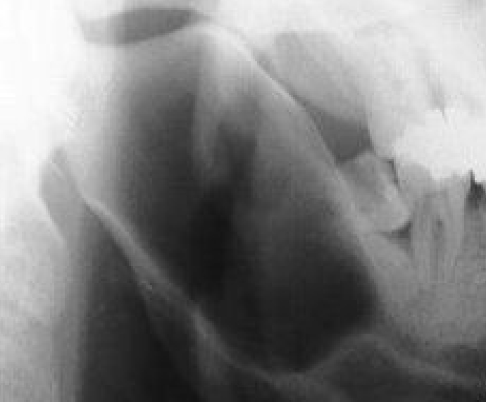
  • Neurofibroma – may present as widening of inferior alveolar canal (Figure 1).
  • Figure 1. Oblique lateral radiograph of the right mandible, showing a well-defined corticated radiolucency. The corticated margins are continuous with the ID canal, suggesting a differential diagnosis of a lesion arising from the contents of the ID canal, in this case a neurofibroma.

    Metabolic disorders include:

  • Osteoporosis;
  • Osteomalacia;
  • Renal osteodystrophy;
  • Osteitis fibrosa cystica (hyperparathyroidism).
  • All cause a loss in bone density making radiographs appear over-exposed and there is also loss of corticomedullary differentiation (loss of lamina dura, outline of maxillary antrum, ID canal tramlines).

    Neoplastic lesions include:

  • Malignant tumours: squamous cell carcinomas (invading mainly from mouth or antrum), osteosarcomas (a symmetrically widened periodontal membrane in a single tooth may be earliest indication), lymphomas (ill-defined lesions), and multiple myeloma (‘punched-out’ ovoid lesions).
  • Metastases: from prostate, thyroid, kidney, lung and breast tumours (but 30% originate from an occult primary lesion) typically have ill-defined borders. Posterior mandibular metastases are four times more common than maxillary.
  • Radio-opacities

    Radio-opaque lesions may be either of soft tissue origin or bony origin and therefore require localization with radiographs taken from two different aspects, and include:

  • Unerupted teeth;
  • Foreign bodies;
  • Calcified lymph nodes;
  • Salivary stones.
  • Congenital and developmental anomalies, such as torus and other bone lumps. Gardner's syndrome (colorectal polyposis, soft-tissue tumours, and skeletal abnormalities) an autosomal dominant condition caused by adenomatous polyposis coli (APC) gene mutation, with multiple osteomas and often impacted and supernumerary teeth and odontomas. Carriers may also have jaw radio-opacities.
  • Odontogenic cysts and tumours (Articles 15 and 16).
  • Fibro-osseous lesions.
  • Inflamed and infected lesions:
  • Odontogenic infections
  • Osteomyelitis – shows no imaging findings until the acute inflammatory reaction leads to bone lysis (osteolysis). Bone density has to fall by 30–50% to show on plain radiography and this usually takes 2–3 weeks (Figure 2). Plain radiographs, and more accurately CT (either MSCT [multislice CT] or CBCT [Cone Beam]) can demonstrate the osteopenia and cortical lysis (including the inferior alveolar canal and mental foramen), sequestra and periosteal new bone formation. MRI has high sensitivity in detecting cancellous marrow abnormalities. In osteomyelitis, periosteal new bone apposition causes cortical thickening and mandibular enlargement, most common on the buccal plate of the mandibular angle or body, especially in young people. Radiologically this may have a lamellar ‘onion skin’ appearance. Swelling of masseter and medial pterygoid muscles is common and both CT and MRI show soft-tissue inflammation, especially in the masticatory and submandibular spaces.
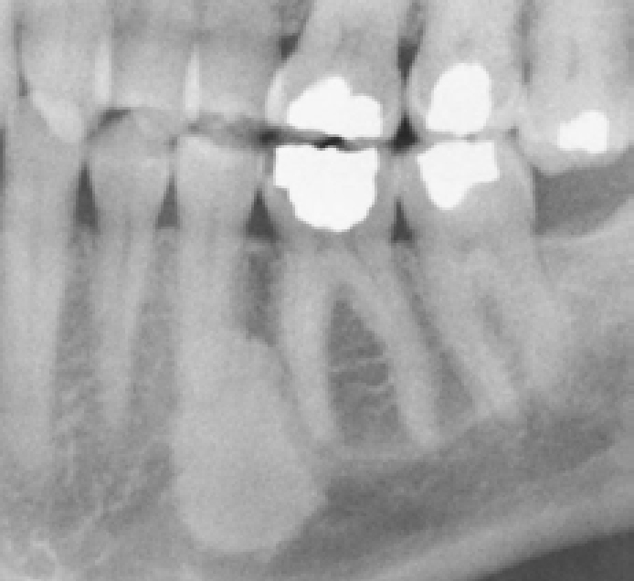
  • Idiopathic osteosclerosis (dense bone island) – area of dense bone in the jaw without apparent cause or signs or symptoms, typically seen in the mandibular premolar/molar area, which may be associated with root resorption (Figure 3).
  • Osteonecrosis. This may follow radiation (osteoradionecrosis; ORN) or drug use (bisphosphonate-related osteochemonecrosis of the jaws; BRONJ).
  • Figure 2. DPT, showing ill-defined radiolucency and radio-opacity within the left body of the mandible. Note the loss of the lower cortex and ID canal tramlines compared to the right side. This was a case of osteomyelitis.
    Figure 3. Section of a DPT showing well-defined increased density of alveolar bone associated with no obvious clinical or radiological pathology, diagnosed as idiopathic osteosclerosis (dense bone island).

    Primary non-odontogenic tumours, eg prostatic carcinoma and breast metastases are occasionally radio-opaque. Sarcomas can cause osteolytic or osteoblastic lesions.

    Mixed radiolucent and radio-opaque lesions

    Mixed radiolucent and radio-opaque lesions are mainly fibro-osseous lesions, inflammatory processes (eg osteomyelitis, actinomycosis, osteonecrosis) and, less commonly, odontogenic tumours (mainly adenomatoid odontogenic tumour and calcifying epithelial odontogenic tumour). Florid osseous dysplasia is one of the commonest causes of multiple radio-opacities of the tooth-bearing area of the jaws.