References
Stafne's bone cavity – unusual presentation in the anterior mandible
From Volume 45, Issue 4, April 2018 | Pages 340-341
Article

Case report
A 55-year-old male was referred to the Oral and Maxillofacial Surgery (OMFS) Department at St George's Hospital by his general dental practitioner for further investigations of an incidental finding of a radiolucency of the anterior mandible. The patient's medical history was unremarkable; he was a non-smoker and revealed no history of previous facial trauma or surgery. Initial examination revealed a moderately restored adult dentition with average oral hygiene. There was neither pain nor swelling of the buccal and lingual sulci of the mandible, and no cervical lymphadenopathy. All teeth were vital, non-mobile, not tender to percussion and no caries was identified.
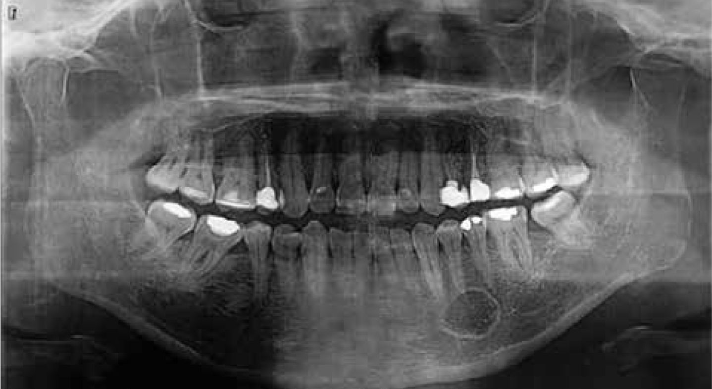
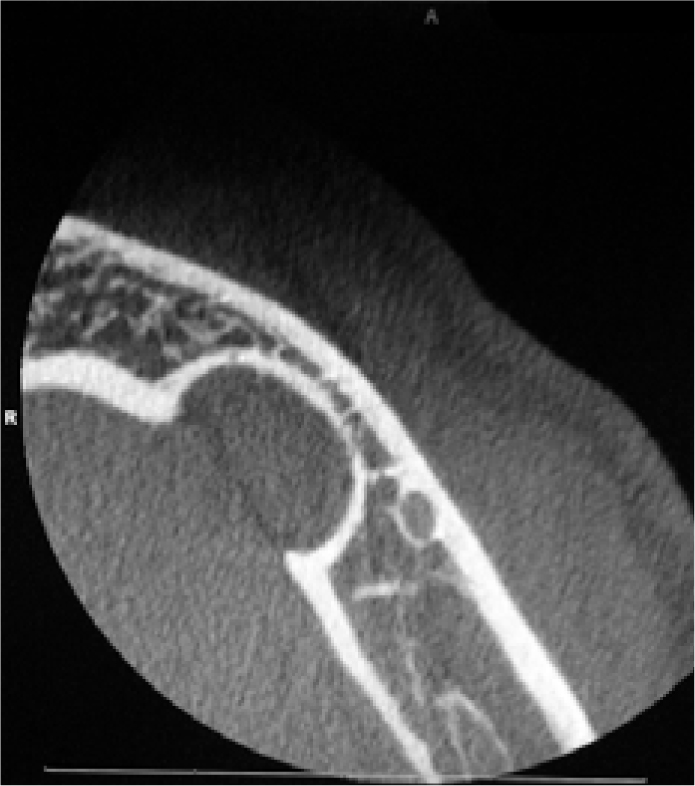
An orthopantomogram (OPG) showed a well-defined, unilocular radiolucency below the apices of an unrestored and non-carious lower left first premolar (LL4). It was decided that a cone beam computed tomogram (CBCT) would be beneficial in diagnosing this lesion and in treatment planning; three-dimensional views allow an accurate assessment of size, location, and proximity to vital structures. Although not a routine imaging modality for SBC, we decided that, as we did not have a definitive diagnosis of SBC from the OPG, it would be necessary should any surgical intervention be required (Figures 1 and 2).


The case was discussed in the OMFS radiology-pathology multidisciplinary team meeting; it was agreed that a biopsy was not warranted and the patient was discharged back to his dentist.
Literature review
Stafne's bone cavity was originally described by EC Stafne in 1942 and has a reported incidence rate of approximately 0.009% to 0.3%.1,2 The majority of cases are located between the first mandibular molar and the angle of the mandible inferior to the inferior dental canal. A review of the literature revealed under 60 reported cases in the anterior mandible.3
Usually an asymptomatic lesion, it is best described as a depression of the lingual aspect of the mandibular bone. However, its radiographic features may mimic that of an intra-bony cystic or neoplastic lesion. It is most likely to occur as a unilateral lesion in males (M:F 3:1) over the age of 50, however, bilateral cases have been reported.3,4 Specifically with regard to cases of anterior SBC, a wide range (18 to 64 years) with a mean of 43 years was noted.5
SBC is primarily identified on routine dental radiography and presents as a well-defined, unilocular, circular radiolucency; however, diagnosis with plain film alone cannot be accurately achieved.5 Cross-sectional imaging, such as CT or CBCT, are useful. MRI has been advocated due to its ability to confirm the presence of salivary gland tissue within the lesion and to limit the radiographic exposure to the patient.6
Histopathological examination is seldom necessary; however, the cavities are usually filled with normal salivary gland tissue arising from the submandibular gland. There have been reports of adipose tissue, blood vessels, skeletal muscle, fibrous connective tissue or lymphoid tissue herniating into the space.2,3
Several pathogenic theories have been suggested in relation to SBC, but the congenital malformation theory (which suggests salivary gland tissue gets entrapped within the mandible during development) is the most universally accepted.6 Lesions as a result of congenital malformation are typically associated with a thin but intact lingual cortex, but this is not consistent with our case.7 Another theory is that of adjacent structures, such as the submandibular gland compressing the lingual aspect of the mandible or local lymphatic infiltration, which may be the causative factor in our case.8
Conclusion
This is another case which highlights that SBC can present at any position along the mandible. There are multiple differential diagnoses for radiolucent lesions found on imaging. We aim to increase the awareness amongst dental practitioners as a conservative management approach is warranted. This will help to prevent unnecessary dental or surgical intervention, therefore improving care and decreasing the morbidity to patients.
