Lin S, Pilosof N, Karawani M Occurrence and timing of complications following traumatic dental injuries: a retrospective study in a dental trauma department. J Clin Exp Dent. 2016; 8:e429-e436 https://doi.org/10.4317/jced.53022
Smith R, McColl E, Bryce G Top tips for managing enamel infractions, cracks and fractures – part 1: diagnosis. Br Dent J. 2023; 234:787-790 https://doi.org/10.1038/s41415-023-5984-5
Lauridsen E, Hermann NV, Gerds TA Combination injuries 1. The risk of pulp necrosis in permanent teeth with concussion injuries and concomitant crown fractures. Dent Traumatol. 2012; 28:364-370 https://doi.org/10.1111/j.1600-9657.2011.01102.x
Lauridsen E, Hermann NV, Gerds TA, Ahrensburg SS, Kreiborg S, Andreasen JO Combination injuries 2. The risk of pulp necrosis in permanent teeth with subluxation injuries and concomitant crown fractures. Dent Traumatol. 2012; 28:371-378 https://doi.org/10.1111/j.1600-9657.2011.01101.x
Lauridsen E, Hermann NV, Gerds TA, Ahrensburg SS, Kreiborg S, Andreasen JO Combination injuries 3. The risk of pulp necrosis in permanent teeth with extrusion or lateral luxation and concomitant crown fractures without pulp exposure. Dent Traumatol. 2012; 28:379-385 https://doi.org/10.1111/j.1600-9657.2011.01100.x
Andreasen FM Transient apical breakdown and its relation to color and sensibility changes after luxation injuries to teeth. Endod Dent Traumatol. 1986; 2:9-19 https://doi.org/10.1111/j.1600-9657.1986.tb00118.x
Bratteberg M, Thelen DS, Klock KS, Bårdsen A Traumatic dental injuries and pulp sequelae in an adolescent population. Dent Traumatol. 2021; 37:294-301 https://doi.org/10.1111/edt.12635
Bourguignon C, Cohenca N, Lauridsen E International Association of Dental Traumatology guidelines for the management of traumatic dental injuries: 1. Fractures and luxations. Dent Traumatol. 2020; 36:314-330 https://doi.org/10.1111/edt.12578
Fouad AF, Abbott PV, Tsilingaridis G International Association of Dental Traumatology guidelines for the management of traumatic dental injuries: 2. Avulsion of permanent teeth. Dent Traumatol. 2020; 36:331-342 https://doi.org/10.1111/edt.12573
Day PF, Flores MT, O'Connell AC International Association of Dental Traumatology guidelines for the management of traumatic dental injuries: 3. Injuries in the primary dentition. Dent Traumatol. 2020; 36:343-359 https://doi.org/10.1111/edt.12576
Levin L, Day PF, Hicks L International Association of Dental Traumatology guidelines for the management of traumatic dental injuries: General introduction. Dent Traumatol. 2020; 36:309-313 https://doi.org/10.1111/edt.12574
Walker L, Pandya J-K, Fletcher S Dental resorption. From diagnosis to management: an update for the GDP. Dent Update. 2024; 51:653-656 https://doi.org/10.12968/denu.2024.51.9.653
Alghaithy RA, Qualtrough AJ Pulp sensibility and vitality tests for diagnosing pulpal health in permanent teeth: a critical review. Int Endod J. 2017; 50:135-142 https://doi.org/10.1111/iej.12611
Bastos JV, Goulart EM, de Souza Côrtes MI Pulpal response to sensibility tests after traumatic dental injuries in permanent teeth. Dent Traumatol. 2014; 30:188-192 https://doi.org/10.1111/edt.12074
D'Souza JL, Mala K, Grover S, Singh A Navigating the digital frontier: transforming endodontic diagnosis through digitization. Part 1. Dent Update. 2024; 51:720-728 https://doi.org/10.12968/denu.2024.51.10.720
Van der Vyver PJ, Vorster M, Jonker CH, Potgeiter N Calcific metamorphosis – a review of literature and clinical management. S Afr Dent J. 2020; 75:316-322 https://doi.org/10.17159/2519-0105/2020/v75no6a5
Patel S, Saberi N, Pimental T, Teng PH Present status and future directions: Root resorption. Int Endod J. 2022; 55:892-921 https://doi.org/10.1111/iej.13715
West JD The aesthetic and endodontic dilemmas of calcific metamorphosis. Pract Periodontics Aesthet Dent. 1997; 9:289-293
Siddiqui SH, Mohamed AN Calcific metamorphosis: a review. Int J Health Sci (Qassim). 2016; 10:437-442 https://doi.org/10.12816/0048738
Oginni AO, Adekoya-Sofowora CA Pulpal sequelae after trauma to anterior teeth among adult Nigerian dental patients. BMC Oral Health. 2007; 7 https://doi.org/10.1186/1472-6831-7-11
Vinagre A, Castanheira C, Messias A, Palma PJ, Ramos JC Management of pulp canal obliteration – systematic review of case reports. Medicina (Kaunas). 2021; 57 https://doi.org/10.3390/medicina57111237
Braga Diniz JM, Diniz Oliveira HF, Pinto Coelho RC Guided endodontic approach in teeth with pulp canal obliteration and previous iatrogenic deviation: a case series. Iran Endod J. 2022; 17:78-84 https://doi.org/10.22037/iej.v17i2.36830
Smith R, McColl E, Bryce G Top tips for restoration of root-filled teeth: Part 1 – minimally invasive techniques for anterior teeth. Br Dent J. 2022; 233:592-594 https://doi.org/10.1038/s41415-022-5168-8
Al-Manei KK, Alzaidi S, Almalki G, Al-Manei K, Almotairy N Incidence and influential factors in pulp necrosis and periapical pathosis following indirect restorations: a systematic review and meta-analysis. BMC Oral Health. 2023; 23 https://doi.org/10.1186/s12903-023-02826-1
Zarow M, Ramírez-Sebastià A, Paolone G A new classification system for the restoration of root filled teeth. Int Endod J. 2018; 51:318-334 https://doi.org/10.1111/iej.12847
Yamashita FC, Previdelli ITS, Pavan NNO, Endo MS Retrospective study on sequelae in traumatized permanent teeth. Eur J Dent. 2017; 11:275-280 https://doi.org/10.4103/ejd.ejd_85_17
This article uses a series of clinical case examples to highlight the importance of comprehensive history taking and examination when carrying out a dental inspection of a patient with an unrestored discoloured anterior tooth. The role of dental trauma in tooth discolouration and advice on clinical intervention is discussed.
CPD/Clinical Relevance: Management of patients with unrestored discoloured anterior teeth may be optimized by early identification and diagnosis of the problem.
Article
The unrestored discoloured anterior tooth (UDAT) is a relatively common finding in general dental practice. Colour changes may be subtle, with insidious onset, and some patients may be both unaware their tooth is discoloured and unable to recollect any precipitating factors.
It is not uncommon for the unconcerned and asymptomatic patient to have been seen by multiple dental professionals over the years without full investigation of their UDAT. However, discoloured teeth should ideally be identified and assessed during routine dental inspection and, where such teeth have been noted previously, legacy records should be consulted to determine if and how the situation has changed.
Depending on the diagnosis, appraisal of treatment options and patient wishes, with consent, active monitoring or restorative/surgical intervention may be planned for a UDAT.
This article outlines the aetiology of single-tooth discolouration and presents the results of a service evaluation of UDAT in primary care, illustrated with case examples. The authors also offer advice on history taking, clinical and radiographic examination, and management of these teeth.
Aetiology
The optical properties of a tooth are modulated by the composition and thickness of the enamel, dentine and pulp, which in turn are influenced by the age of these tissues and their relative health or disease status.
Pathological states that may contribute to the (dis)colouration of a tooth include developmental disease, caries, cracks, fractures, pulpal injury (and its various sequelae such as pulpal haemorrhage and haemolysis, calcific metamorphosis and necrosis with or without endodontic infection), and internal coronal or external cervical resorption.
A tooth may experience many pathological insults over time, with associated changes in colour. For example, a traumatic dental injury (TDI) may result in cracks that can appear brown owing to ingress of both bacteria and extrinsic stains (internalized discolouration).1
In conjunction with any disruption to pulpal blood supply, cracks that extend deeper into the tooth structure may enable bacteria to drive low-grade inflammation within the pulp tissue, potentially stimulating progressive calcific metamorphosis (yellow discolouration) or pulp necrosis (grey discolouration).
The discolouration of a single tooth in the absence of caries or a deep restoration should raise suspicion of previous physical injury. TDIs may arise via excessive forces, applied via a fleeting impact (such as blunt force trauma during an accident or assault) or over prolonged time periods (via excessive forces during orthodontic treatment). Parafunctional habits, such as bruxism, may also play their part.
In addition, the (dis)colouration of a tooth will be modified by the presence and nature of any surface stains. This tends to be a more generalized problem, and is not discussed further here.
The annual global incidence of dental trauma is estimated to be up to 4.5%, although this varies considerably depending on country and culture.2 Up to 25% of adolescents/young adults will receive at least one TDI, with a higher risk to upper anterior teeth with an increased overjet, and in those leading more active lifestyles and taking part in contact sports.2 However, many of these incidents will be minor and result in the lower end of severity and damage. As a result, it is estimated that only one-third present to a dentist within 24 hours, with the other two-thirds delaying for up to 1 year.3 The true incidence of TDIs is therefore undoubtedly under-reported.
This means that many who receive a minor knock to a front tooth never report this to their dentist, with the majority experiencing no long-term sequelae, and it is suspected that the patient eventually forgets about such incidents. However, calcific metamorphosis is common after concussion and luxation injuries (up to 35%), resulting in gradual discolouration of the tooth over many years, even though the patient may be completely unaware of any problems.4
The general relationship between specific key findings of diagnoses and tooth discolouration is summarized in Table 1.
Key finding/diagnosis
Definition
Effect on tooth (dis)colouration
Crack
Defect where there is a break between two parts of a tooth, without separation of the fragments. May involve dentine +/- enamel +/- cementum +/- direct communication with pulp5
Possibly none, but discolouration may arise from ingress of bacteria and extrinsic stain into the crack, which may become light orange/brown or, occasionally, blackModulating factors: location/number/depth of defect(s), and presence and nature of extrinsic staining agents, e.g. bacterial species, tobacco and dietary sources
Fracture
Defect where there is a break between two parts of a tooth and the fragments have separated. May involve one or more of the following hard tissues: enamel, dentine or cementum, and direct communication with pulp5
Non-cleansable planes of fracture will accrue biofilm and extrinsic stains and become brown or blackModulating factors: as for crack.Combination of fracture and luxation injuries will have a negative synergistic effect on outcome6,7,8
Calcific metamorphosis
A pulpal response to trauma characterized by an increased rate of deposition of hard tissue within the pulp chamber/root canal space.This can occur in up to 35% of teeth that have received a traumatic dental injury. Progressive partial calcification may ultimately result in the radiographic evidence of complete obliteration of the pulp chamber and root canal. Histological sections of such teeth usually demonstrate vestiges of remaining pulp space9
Yellow hue in early stages, becoming dark yellow/orange/light brown over time as more hard tissue is depositedModulating factors: location/density of calcification within pulp tissue, pulp status (vital/non-vital with or without endodontic infection)
Coronal resorption
Resorption is a condition associated with a physiologic or a pathologic process resulting in loss of dentine, cementum and/or bone.9Internal coronal resorption and external cervical resorption may both result in discolouration of the crown. Root resorption can also occur following minor trauma but does not affect coronal colour
Localized pink spot within the gingival-half of the clinical crown – an uncommon presentationModulating factors: extent of the resorption lesion and pulp status
Pulpal haemorrhage
Rupture of intra-pulpal blood vessel(s). Blood may be hydraulically driven into the coronal dentinal tubules. The extravasated blood then breaks down (e.g. haemolysis) releasing haemoglobin, which may then be metabolized into haemosiderin and bilirubin9
Initially pink or red hue (which in the presence of a vital pulp can occasionally resolve spontaneously), though more commonly further darkening (to a purple/brown) takes place due to formation and persistence of haemolytic products10Modulating factors: extent of haemorrhage, pulp status (vital/non-vital with or without endodontic infection)
Pulp necrosis
Death of the dental pulp. Following a traumatic dental injury, 7–34% of patients may develop pulp necrosis.4,11 Of teeth with calcific metamorphosis 7–27% may develop pulp necrosis with signs of chronic apical periodontitis, and this combination provides a particular clinical challenge12, 13
Grey due to pulp breakdown products created via bacterial action. Sometimes a tooth may appear black if black pigmented anaerobes are presentModulating factors: nature of endodontic infection
Service evaluation of management
Triggered by curiosity, the lead author conducted a service evaluation to assess the management of UDAT identified in military patients presenting to him for routine dental inspection over a 32-month period. The service evaluation was registered within Defence Primary Healthcare's audit register.
Patients identified with UDAT had their history taken alongside a retrospective review of their clinical records to assess whether the UDAT had previously been identified and investigated.
The clinical examination involved pulp sensibility testing of affected teeth using both cold (Roeko Endo-Frost, Coltène Whaledent, Germany) and electrical pulp testing (Digitest 3, Parkell, New York, USA). Clinical photographs and long cone peri-apical (LCPA) radiographs were taken using a direct digital imaging sensor with evaluation using VixWin Platinum software (Gendex Dental Systems, PA, USA).
Clinical management depended on the underlying aetiology of the UDAT and patient preference. Patient consent was given for the storage of the pertinent findings in an Excel database (Microsoft, WA, USA) and for the use of clinical photographs and copies of radiographs.
Results
During the study period, 21 discoloured incisors (12 upper and six lower centrals; 86%), and one upper and two lower laterals (14%) were identified in 19 patients. Only nine patients (47%) could recall an episode of previous dental trauma as a possible cause for their discoloured tooth; these included significant events such as assaults, sports injuries, a traffic accident and a swimming pool incident.
Fourteen of the 21 teeth (67%) had received no previous radiograph, and 15 had not been subjected to any previous sensibility tests (71%). Of the 14 UDATs with no previous radiograph, five were found to have undiagnosed apical disease (36%), and one was found to have a root fracture (~7%). One of the teeth, discovered to have a large apical radiolucency, was a non-discoloured lateral incisor adjacent to a UDAT (Case 5).
Twelve teeth (57%) required further monitoring only, eight required endodontic treatment (38%) and one required extraction and prosthetic replacement (~5%).
This indicates a significant underdiagnosis of dental morbidity, and serves to underline the importance of applying assessment and monitoring guidelines for traumatized teeth as set out by the International Association of Dental Traumatology, which maximize the probability of achieving the most favourable outcome.14,15,16,17,18 Findings from the service evaluation are summarized in Table 2 and a selection of representative cases is shown in Table 3.
Case
Patient demographics
History
Key diagnoses following clinical and radiographic assessment
Dental trauma via impact with TV remote control (thrown by brother) 25 years ago
4
No
No
Mid-third horizontal root fracture Calcific metamorphosis Replacement resorption of apical fragment UR1
Referred for extraction and prosthetic replacement UR1 with implant-supported crown
16
43
Fit and well
Non-smoker
6
LL1
Yes
Punched in face 13 years ago
3
Yes (EF only)
Yes
Calcific metamorphosis LL1
Clinical and radiographic monitoring
17
43
Fit and well
Non-smoker
4
UL1
Yes
Dental trauma during rugby game as a teenager
4
No
No
Enamel hypoplasia and calcific metamorphosis UL1
Clinical and radiographic monitoring
18
35
Fit and well well
Non-smoker
8
UL1
No recollection
–
4
No
No
Calcific metamorphosis UL1
Clinical and radiographic monitoring.
19
23
Fit and well
Smoker
4
UL1
No recollection
–
4
No
No
Calcific metamorphosis UL1
Clinical and radiographic monitoring. External bleaching
DMFT: decayed, missing and filled teeth; EF: Endo-Frost cold test; EPT: electric pulp test; LCPA: long cone peri-apical; RCT: root canal treatment; UDAT: unrestored discoloured anterior tooth.
*Dental inspections within the 5-year period before attendance with the first author, or since date of trauma (if known and earlier).
Service evaluation case reference
Key findings and diagnoses
Clinical appearance
Radiographic appearance
Treatment options included
8
Orange/light brown discolouration UL1 Partial calcific metamorphosis, secondary to tooth wear (modified by abrasion, erosion and attrition) Peri-apical tissues are healthy
Preventive with or without restorative management of tooth wear Long-term clinical and radiographic monitoring with or without external bleaching
9
Yellow/ochre discolouration UR1 Complete calcific metamorphosis. Replacement resorption of apical half of root Likely to be ankylosed19
Long-term clinical and radiographic monitoring with or without external bleaching. The red line on the radiograph is a measurement tool that enables the amount of root lost to resorption to be estimated
6
Brown discolouration. Attritive type incisal tip tooth wear (with dentine exposure). Vital pulp was likely (tooth tested positive with Endo-Frost and EPT). Peri-apical tissues are healthy. Discolouration possibly due to increased peri-tubular dentine deposition
Long-term clinical and radiographic monitoring with or without external bleaching Note: RCT is likely to be challenging owing to the 1–2 root canal configuration (bifurcating in the mid-third of root)
14
Yellow-brown discolouration LL1 Complete calcific metamorphosis Peri-apical tissues are healthy The more intense colour demonstrates the variations in hue, compared to Case 9, that may be seen in cases of long-standing complete calcific change
Long-term clinical and radiographic monitoring with or without external bleaching If the latter is chosen, because of the advanced nature of the calcific metamorphosis, the patient should be advised that resolution to the original shade may be difficult
15
Light brown discolouration UR1. Mid-third root fracture, with apparent bony healing and calcific metamorphosis of coronal portion, and replacement resorption of the apical fragment
Long-term clinical and radiographic monitoring with or without external bleaching Or, extraction and prosthetic replacement (denture/bridge/implant)
10
Grey discolouration UR1 Pulp necrosis. Radiographic evidence of early peri-apical disease
RCT and inside–out bleaching, with a porcelain veneer should bleaching not be fully successful and the patient still concerned about the colour
Pink hue in cervical third UR1 (suggestive of pulpal haemorrhage following recent trauma). Enamel infraction lines. Periodontal ligament widening, which from case history, likely to be associated with recent luxation-type injury.
Long-term clinical and radiographic monitoring and endodontic/restorative intervention as required over time
History taking and clinical examination
A full and robust history should be taken for any patient presenting with a UDAT, followed by thorough extra-oral and intra-oral examination.
Specifically, history taking should screen for:
Patient awareness of discoloured tooth/teeth, onset of discolouration, related symptoms (e.g. pain/swelling),
Recollection of any dental trauma, including its nature, timing and any previous treatment delivered in relation to this (e.g. tooth repositioning/splinting),
Details of any previous orthodontic treatment (if applicable),
Any restorative treatments (including external bleaching and resin infiltration).
The examination should include:
Assessment of the character of (dis) colouration, as this often points to its aetiology (Table 1);
Recording of the discolouration (ideally in conjunction with a shade guide and digital photography);
Tenderness to palpation of the tooth itself and adjacent alveolar mucosa;
Tenderness to percussion; mobility; relevant occlusal factors (e.g. unfavourable guidance with or without overloading/fremitus);
Presence of caries or cracks (with or without transillumination);
Status of any restorations;
Six-point periodontal charting.
Given the importance of special tests in ascertaining the diagnosis, sensibility testing and radiographic imaging are discussed in greater detail.
Pulp sensibility testing
Sensibility is the ‘capacity for sensation or feeling; responsiveness or susceptibility to sensory stimuli’.20
Sensibility testing of teeth provides a surrogate measure of pulpal health, estimated by the knowledge that an intact nerve supply (indicated by a positive sensibility test) correlates to the presence of an intact vascular supply (e.g. vital pulp). However, this correlation is imperfect, and there is the possibility of both false negative and false positive results, particularly if a TDI has been experienced within the past 12 months.21,22
The most common sensibility tests used in dental practice are cold testing (e.g. using a refrigerant, such as Endo-Frost (Coltène Whaledent), sprayed onto a cotton wool pledget and held on a tooth with tweezers), and electric pulp testing (EPT). Pulp sensibility test results are recorded as ‘positive’ or ‘negative’.
Additionally, as contemporary EPT devices provide arbitrary numbers to indicate the amount of current flow, this can be recorded in the clinical notes for reference at follow-up as required. However, the clinician should not put too much emphasis on the number itself, as variations between EPT devices (if more than one is used in practice), electrode positioning on tooth, the nature of electrolyte medium (if used), as well as modulation of the psychological state of the patient will influence the point at which they will indicate a positive test. Reasons for false negative results include, but are not limited to:
Under-developed neural networks in immature teeth;
TDI resulting in direct damage to the neural supply;
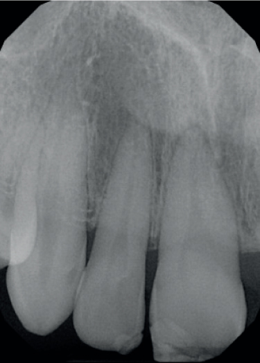
Calcific metamorphosis (see below), where the additional dentine thickness acts as both a thermal (Figure 1) and an electrical insulator.14,22
Figure 1. Radiographic series (from an unrestored discoloured anterior tooth case not included in the service evaluation) to illustrate suspected pulpal misdiagnosis in an UL1. Clinically, the tooth was asymptomatic, but had a yellow discolouration. Radiographically, there was evidence of calcific metamorphosis in the crown and apparent peri-apical health. The attending GDP made a diagnosis of pulp necrosis UL1, principally on the basis of a negative cold test (an electric pulp test (EPT) was not conducted) and root canal treatment (RCT) was initiated. Unfortunately, the attempted endodontic access was misaligned. The GDP could not locate the root canal and, inadvertently, perforated subgingivally through the buccal aspect of the root. The GDP then referred the patient for specialist management, whereupon the UL1 was found to be asymptomatic, testing positive with EPT and, at that point, still had no radiographic signs of apical pathosis; therefore, the tooth was scheduled for external bleaching only. However, upon review 4 months later, the UL1 tooth was found to have developed chronic apical periodontitis, with the lesion extending over the apex of the UL2 (which was not discoloured and tested positive with Endo-Frost (Coltène Whaledent) and EPT). The UL1 was treated definitively with RCT, the perforation was repaired with Biodentine (Septodont, Saint-Maur-des-Fossés, France), and inside/outside bleaching was performed. Peri-apical healing was demonstrated at follow-up, and the UL2 continued to test positive with pulp testing. This case demonstrates both the value of pulp testing with cold and EPT, as well as highlighting the potential challenge of endodontic access involving a tooth with calcific metamorphosis.
Reasons for false positive results include, but are not limited to:
The persistence of functioning nociceptor fibres within dying or necrotic pulp;
Transmission of stimulus to nociceptors within the periodontal ligament via fluid in crown-root cracks or fractures in a non-vital tooth;
Inadvertent stimulation of the pulp in an adjacent tooth.
It should be noted that while a positive response shortly after a TDI is considered a good predictor of pulp survival, a negative response is not necessarily associated with subsequent pulp necrosis.22
Despite some disadvantages, EPTs are still a valuable resource to clinicians and their use in cases of UDAT is recommended to aid diagnosis and to provide a baseline reference against which to monitor any change over time.
True vitality tests, such as laser Doppler flowmetry and pulse oximetry, have been used in isolated cases to assess revascularization and changes in pulp microcirculation of traumatized teeth. Both technologies have yet to be refined to the point of application in real-world dentistry, but the latter in particular offers a promising objective assessment of arterial blood flow that, with more research, might be beneficial in future dental practice.23
Radiography
The initial radiographic examination of UDAT should be with LCPA images. When reporting the radiograph(s), the clinician should ideally consider:
Teeth present;
Restorative status;
Crestal bone levels;
Pulp chamber/root canal anatomy (e.g. canal(s) present, and whether the size of canal(s) is commensurate with patient age/adjacent teeth or if abnormally narrowed/obliterated (owing to calcific metamorphosis) or enlarged (because of internal resorption);
If the periodontal ligament space is within normal limits (or not);
Peri-radicular pathosis;
The presence/type of any external root resorption.
If the information discernible from LCPA radiograph(s) is insufficient for accurate diagnosis, prognostication or treatment planning, 3D imaging via CBCT is advised.24
Diagnosis
Following history-taking and clinical/radiographic examination, information is synthesized and definitive (or provisional) diagnoses are formulated. Diagnosis statements for UDAT will often mention discolouration (type), cracks/fractures in the crown/root (if applicable), the presence of resorption (if applicable) and endodontic and periodontal status.
When to intervene
It is beyond the scope of this article to categorize and provide detailed management advice for every TDI or resorption variant; comprehensive guidance on these topics has been published elsewhere.14,15,16,24,25 However, in Table 4, the authors offer a summary of suggested clinical situations for when intervention for the UDAT may be justified.
While the rationale for endodontic treatment of teeth with symptoms, frank peri-apical disease or progressive resorption is relatively clear, the pros and cons of intervention in cases of the more common sequelae of calcific metamorphosis can often provoke debate.27
Up to 75% of calcific metamorphosis cases are symptom free and have no apical pathology, so require no treatment other than radiographic review.12 If monitored in this way then, unless rapidly progressive, prophylactic treatment of calcific metamorphosis ‘does not seem to be justified’, ‘should only be considered in rare circumstances’ or is even ‘contraindicated’.24,28,29 The risks of endodontic intervention in asymptomatic cases without apical radiolucency may often outweigh any perceived benefits, as shown in Figure 1.
How to intervene
Ideally, the patient should be fully informed of the rationale and relative advantages and disadvantages of different treatment approaches to support their decision-making and enable the consent process.
While there are multiple options for treating UDAT, a minimally invasive approach should always be considered in the first instance. This includes:
Clinical and radiographic monitoring.
Single tooth external bleaching via either bleaching tray or in-office technique. Following bleaching, extrinsic stains (via crack lines), may (again) penetrate the tooth structure and cause the tooth to discolour. To reduce the risk of this, cracked surfaces may be sealed with a layer of composite resin (or resin infiltration) following bleaching;
Direct composite veneer: underlying discolouration is likely to shine through with standard translucent materials and, without prior bleaching or making the composite layer particularly thick, this approach will often be unsuccessful. It may therefore be necessary to use more opaque composite layers to mask such discolouration and an unbonded trial can quickly determine whether this approach would be of benefit.
Porcelain veneer: unless the porcelain veneer contains an effective opacious layer, it may not be able to mask the underlying discoloured tooth tissue. The addition of the opacious layer creates bulk thickness to the veneer (up to 1 mm in total), and additional tooth preparation will be required.
Root canal treatment and inside-outside bleaching for non-vital teeth: preparation of the access cavity and root canal location/negotiation in teeth with calcific metamorphosis can be particularly challenging. The authors recommend that initial access is completed without a rubber dam in place to aid orientation, via the visualization of both adjacent teeth and the root orientation within the alveolar envelope, and that a rubber dam is placed immediately after this, before the surgical area is disinfected with a cotton wool pledget soaked in sodium hypochlorite. Key anatomical landmarks and colour changes can be used to locate the root canals.30 The use of a dental operating microscope is very beneficial in the treatment of such cases.31,32 The process for inside-outside bleaching is highly technique sensitive and a recommended strategy for this has been previously described.33
Crown: the preparation for a full-veneer restoration is destructive and carries greater risk to the pulp health.34 However, if other strategies have failed to attain the desired aesthetics, or if the tooth is to be included in a more generalized restorative plan, crowning of the tooth may be indicated.
Post/core and crown: following root canal treatment, if the tooth is broken down to the extent that a crown cannot be predictably retained, a post/core is indicated. Where there are at least two axial walls or a <2 mm ferrule is possible, then a cast post is indicated over a fibre type.35
Extraction and prosthetic replacement: this is applicable if the tooth cannot be predictably restored.
Summary
The causes of minor trauma involving anterior teeth are varied, and any sequelae may not manifest for many years, if at all.36 The gradual discolouration of an affected tooth may be subtle, with patients often unaware of the problem.
In the authors' experience, UDATs are frequently not picked up at routine examinations. Possible reasons for this during a busy day in practice may include colour vision deficiency (which affects 8% of men and 0.5% of women), variations in hue, value and chroma changing under differing light sources (metamerism), fatigue and occasional time constraints.37 If extra time is required to carry out more detailed history-taking and further tests, the authors recommend rescheduling the patient for such additional investigations.
Questions that may also help improve the clinical mindset for identifying a UDAT during a whole-mouth examination include:
Why is this tooth a different colour from its neighbours?
How long has this tooth been a different colour?
Does the patient know what caused this tooth discolouration?
What is the suspected condition of the pulp?
Is there any associated apical pathology?
What is the diagnosis?
What is the most appropriate treatment option?
Conclusion
Effective recognition and diagnosis of UDAT enables appropriate treatment and potentially reduces the risk of a patient experiencing a dental emergency.
In the presence of more complex contributing disease, such as calcific metamorphosis plus apical periodontitis, extensive inflammatory root resorption or root fracture, onward referral may be warranted.12,38,39,40