Article
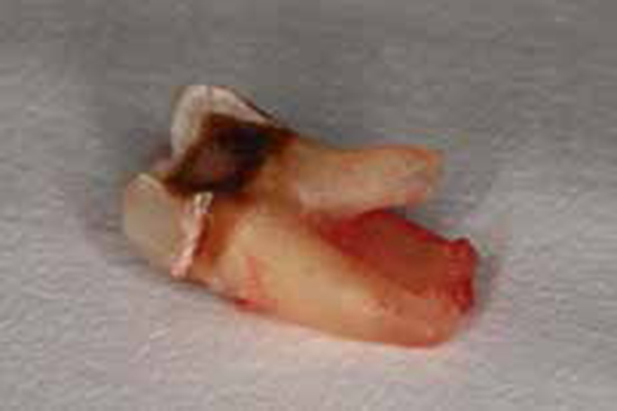
A common complication of exodontia is the fracture of a root tip (Figure 1). Often the root fragment is visible but withdrawing it can be troublesome. It is usually out of reach of conventional surgical instruments, such as luxators and forceps, and longer instruments such as probes cannot grip the fragment sufficiently. While bone removal can be carried out to facilitate access, a quicker and less invasive option has been reported using Hedstrom endodontic files.1

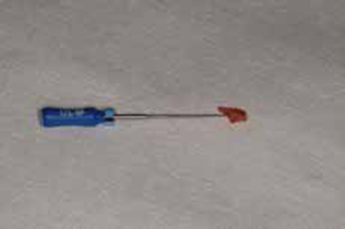
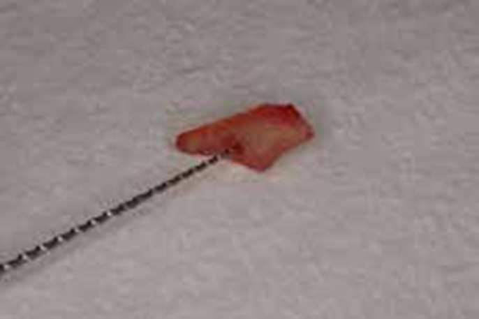
The Hedstrom file is a round stainless steel wire ground to create a sharp-cutting flute. When rotated clockwise this file readily binds to dentine, something to be avoided during normal use in root treatment.2 However, this pitfall can be used to advantage if a root tip fractures. The Hedstrom file can be ‘screwed’ into the root canal of the fractured tip and used to withdraw the fragment (Figures 2 and 3).


Care must be exercised when using this technique. To ensure airway protection, floss or a parachute chain should be attached to the file. The size of the file should correspond to the expected size of the canal, allowing the file to bind firmly. For most teeth around ISO size 30 is likely to be successful. If the file is too large then it will prove difficult to engage into the canal at all, or it might risk fracturing the root tip further if forced into the canal. It is possible to place a large lateral force on a root as the file can have a wedge effect. If the file is too small or not screwed to a sufficient depth, then the file will fail to bind sufficiently to the root. Instrument separation is a potential risk but unlikely, if used with care, as the small piece of root will usually be weaker than the steel instrument.
This technique can only be recommended when the root tip can be visualized in the socket and where the fragment is slightly moveable when gently probed. The root canal of the fragment should be identified to avoid insertion of the file into the periodontal ligament or socket wall. The file can only place a small amount of withdrawing force on the root fragment so, if the anatomy of the root tip is unfavourable, then the file is unlikely to remove the fragment. If the root is close to the maxillary sinus, then particular care must be exercised to avoid excess apical pressure which could displace the fragment. Careful assessment of the pre-operative radiograph, knowledge of anatomy, good operating light, careful fine-tipped suction and magnification are valuable aids.
Consideration should be given to this technique before pursuing more invasive options. Where an immediate surgical approach is appropriate, the technique could still be used to control the position of the fragment while bone removal is carried out, particularly if in proximity to the maxillary sinus or other vulnerable structures.
