Shivakumar A, Bardvalli SG. An alternative approach for re-attachment of the fractured fragment – a case report. Int J Contemp Dent. 2011; 2:(2)48-51
Rajput A, Talwar S, Ataide I Complicated Crown-root fracture treated using reattachment procedure: a single visit technique. Case Reports in Dentistry 2011. 1-5
Oliveira GM, Oliveira GB, Ritter AV. Crown fragment reattachment: report of an extensive case with intra-canal anchorage – case report. Dent Traumatol. 2010; 26:174-181
Altun C, Guven G. Combined technique with glass-fibre reinforced composite post and original fragment in restoration of traumatized anterior teeth – a case report. Dent Traumatol. 2008; 24:e76-e80
Tay FR, Pashley DH. Monoblocks in root canals: a hypothetical or a tangible goal. J Endod. 2007; 33:391-398
‘Richmond crown technique’ – a newer approach for re-attachment TS Ashwini Shivani Shah Salim Makandar Dental Update 2025 40:5, 428-428.
Authors
TSAshwini
Postgraduate Student, Department of Conservative Dentistry and Endodontics, Maratha Mandal's Nathajirao G Halgekar Institute of Dental Sciences, Belgaum, Karnataka, India
Postgraduate Student, Department of Conservative Dentistry and Endodontics, Maratha Mandal's Nathajirao G Halgekar Institute of Dental Sciences, Belgaum, Karnataka, India
Senior Lecturer, Department of Conservative Dentistry and Endodontics, Maratha Mandal's Nathajirao G Halgekar Institute of Dental Sciences, Belgaum, Karnataka, India
Re-attachment is one of the most acceptable techniques followed during the management of complicated coronal tooth fractures, especially when there is an intact fracture fragment. The following technique is a highly conservative, quick, cost-effective approach that combines aesthetics and function, thereby postponing the use of a more aggressive prosthetic solution.1,2
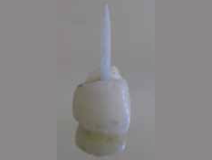
The patient reported with a complicated crown fracture with respect to the maxillary right lateral incisor (UR2) and an uncomplicated fracture to the maxillary right central incisor (UR1) (Figure 1).
Figure 1. Pre-operative photograph of the fractured fragment.
Since the fracture fragment of lateral incisor (UR2) was available, a new approach, the ‘Richmond Crown Technique’ for re-attachment was planned.
Clinical procedure
Local anaesthesia was administered and isolation was achieved.
The fracture fragment was stored in a sterile saline solution to prevent dehydration. Single visit root canal treatment was completed.
The restoration of the tooth in question implied the use of a post system owing to lack of tooth structure coronal to the gingival margin to support the restoration/fragment, thus a post space was prepared to receive a fibre post.3,4
A light transmitting fibre post (Glassix Glass Fibre Composite Post, Nordin, Switzerland) for the desired length and adaptation was obtained.
Preparation of fractured fragment
A box-like preparation was made in the pulp space of the fracture fragment; the remnant of pulp tissue was removed and prepared to receive post and composite.
The box preparation was etched, rinsed and dried; bonding agent (Prime & Bond NT, Dentsply, DeTrey, Germany) was applied onto the inner prepared surface of the fractured fragment and post, which was then cured for 20 seconds each.
Composite (Z250, 3M ESPE, St Paul, MN, USA) and the post were placed into the box and the whole unit was inserted into the canal, checked for approximation of the fractured fragment and cured for 20 seconds.
The whole unit was then withdrawn from the root and then curing was done for an additional 20 seconds. The unit resembled a ‘Richmond Crown’ (Figure 2).
Figure 2. Fractured fragment with post in place, resembling ‘Richmond Crown’.
Attachment of ‘Richmond Crown’ to the root
According to the manufacturer's instructions, the canal was prepared to receive the post.
Resin-luting cement (Rely X Unicem, 3M ESPE) was applied on the post and the whole unit was introduced into the canal.
Excess cement was removed and margins were cured.
The margins were then finished and polished.
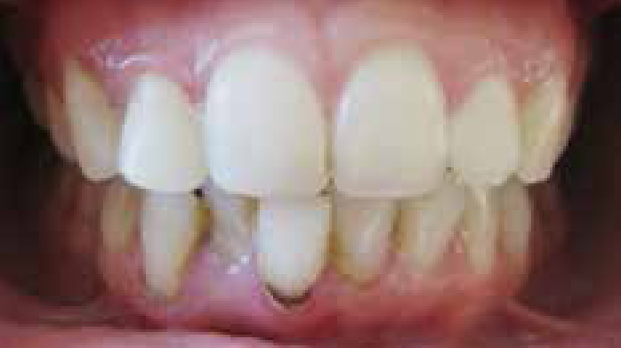
UR1 was restored using composite restorative material (Figure 3).
Figure 3. Post-operative photograph.
Advantages of the ‘Richmond Crown Technique’ for re-attachment:
Less time consuming.
Isolation was less critical since most of the work was done extra-orally.
Monobloc is created, thereby reducing the incidence of failure.5
Composite resin was preferred over resin cement for the attachment of the post onto the fracture fragment (coronal tooth structure) as composites are more colour stable and thus do not discolour over time.1