Article
The Cvek pulpotomy is a useful technique for the management of a complicated crown fracture of vital incisors with open or closed apices. It involves removing 1–3 mm of inflamed pulp, leaving the healthy vital cell-rich pulp to aid healing post trauma. If haemostasis is achieved, the tooth can be dressed and restored to normal function with good prognosis. Miomir Cvek pioneered this approach and found a 96% success rate for teeth treated up to 90 days after trauma with 1–4 mm exposures.1
Trauma may be stressful for patient and clinician alike. Yet this is a simple, inexpensive and relatively quick way of ensuring a tooth has the best prognosis following traumatic injury. It does not involve specialist equipment and uses materials found in everyday general dental practice. This method could be used with reference to February's Technique Tip: ‘Traumatized fractured incisors: re-attachment of the fragment’.2
Case report
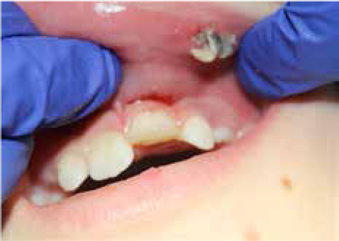
The following case describes a 10-year-old boy with a history of complicated crown fracture to UL1 which he had sustained 48 hours previously. The fractured fragment had been stored in milk since the accident. The UL1 was unreliability negative to sensibility testing with ethyl chloride.
Clinical and radiograph examination revealed (Figures 1, 2, 3):
| 8 | 5 | 1 | 1 | 5 |


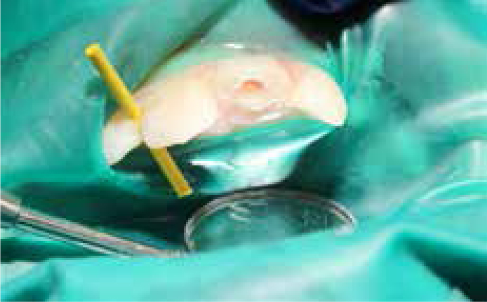
The Cvek pulpotomy


At review two weeks later, the patient reported no pain or discomfort and was happy with the aesthetic results. The UL1 demonstrated good aesthetics and required a 3-month radiograph and clinical review with long-term multidisciplinary assessment for hypodontia and malocclusion.
Discussion
The Cvek pulpotomy can be a useful technique for patients who present up to several days post trauma. It is commonly thought that time between accident and treatment is a crucial factor for success. Cvek, however, found that the time delay from trauma to treatment was not a significant factor. Treatment was carried out between 1 hour and 1260 hours (90 days) after trauma; 96% of those treated within 72 hours survived with no pulpal necrosis. Those treated after 72 hours had a slightly lower prognosis; 87.5% survived with no pulpal necrosis. Even with a delay of treatment, teeth treated with Cvek-type pulpotomy have a good prognosis.1
Cervical or complete pulpotomy is more extensive than the Cvek pulpotomy. It removes the coronal pulp tissue in its entirety to cervical level and places medicament on the canal orifices.3 If more than 3 mm of coronal pulp tissue needs to be removed to control bleeding, a cervical pulpotomy is indicated and this should be done quickly post trauma to ensure the best possible prognosis for the tooth. The clinician cannot know how much pulpal tissue needs amputating until treatment is taking place.4 Materials other than calcium hydroxide have been reported for use for a coronal barrier such as MTA; however, this can result in discoloration around the cervical area.
Maintaining vitality of the immature tooth allows continued growth and hard tissue deposition, which strengthens dentine walls and reduces the risk of cervical fracture.5 It also avoids the resorption and ankyloses which can occur following pulpal necrosis in traumatized immature incisors. For these reasons, and the reported 100% success rate,6 the Cvek pulpotomy is indicated for immature teeth. Some argue that mature teeth, on the other hand, have a diminished healing capacity and should be extirpated following complicated fracture.3 The belief is that once the tooth is fully formed, the pulp has less contact with the periodontium and is limited in the ability to repair itself. From a medico-legal perspective, pulpectomy should not be instigated until signs and/or symptoms are evident. Indeed, the data suggest that mature roots treated with the Cvek pulpotomy have a similar prognosis to immature teeth.
If signs of pulpal necrosis do occur later, there is always the option to extirpate at this stage. The Cvek pulpotomy is, at the very least, a conservative treatment which aims to maintain the nerve of the tooth, allowing the tooth to grow and become a fully functional life-long healthy unit. This is a useful conservative approach which should be considered even if the patient presents several days post trauma.
