Article
The restoration of large Class II cavities with photo-activated composite resin is technically demanding and time consuming and often results in early failure due to sensitivity, secondary caries or tooth substrate fracture. This is often a result of the net volumetric shrinkage of the resin on photo-activation setting up stresses at the restoration-tooth substrate interface giving rise to bond degradation or cuspal fracture, or both. The novel use of an auto-cure glass ionomer cement as a mega-filler completely enclosed by composite resin may offer a strategy to reduce these stresses.
The Super-Closed Sandwich
The Glass Ionomer Composite Super-Closed Sandwich technique was originally described by Magne.1 This is an elegant solution whereby the dentine is initially hybridized with a 4th Generation Bonding Agent (Optibond FL, Kerr, Peterborough, UK). The proximal walls are established with paste composite and pure glass ionomer is injected as a dentine substitute. The occlusal surface is then closed with composite, creating a sealed unit. In this model, the glass ionomer is not bonded to the composite, but rather acts as a ‘Mega-Filler’ particle, reducing polymerization stresses.
This paper aims to present an alternative approach: an autocure Resin-Modified Glass Ionomer (Fuji VIII GP, GC, Newport Pagnell, Bucks, UK). This may offer the advantages of a bond to the overlying composite resin, improved translucency and therefore aesthetics over traditional glass ionomer, and reduced polymerization shrinkage in comparison to photo-activated TriCure Resin-Modified Glass Ionomer Cement (RMGIC). Furthermore, in deeper cavities the hybridized dentine protects the pulp-dentine complex from the hygroscopic effects of the RMGIC. We have termed this approach ‘The Modifed Super-Closed Sandwich Technique’.
A 63-year-old male patient attended the practice complaining of intermittent sharp pain localized to the upper right premolar region with no radiation. The pain was elicited by cold and sweet and was several seconds in duration: it did not disturb sleep.
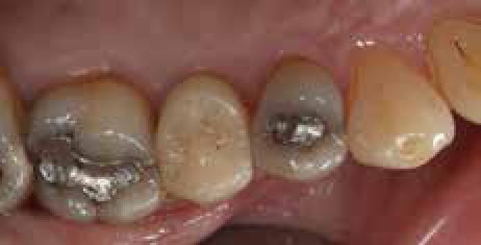
On clinical examination a medium-sized, occlusal amalgam restoration was present in the UR4: a stained crack line was evident mesially extending to an area of visible interproximal discoloured enamel (Figure 1). There were no associated periodontal probing depths: nor was the tooth tender to pressure; electronic and ice pulp testing yielded normal responses.
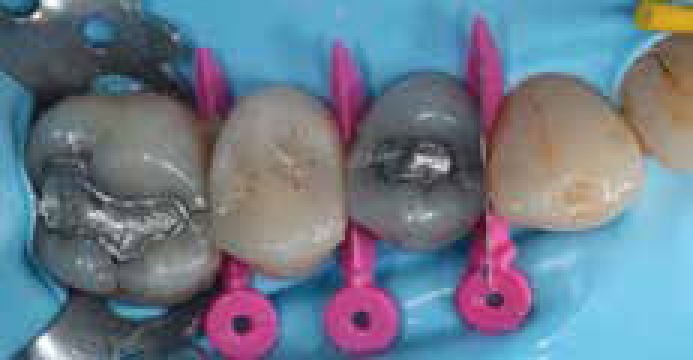
After local anaesthesia, the upper right quadrant was isolated with latex-free rubber dam (D2DEndo) and an Ash 8a rubber dam clamp. Interproximal isolation was achieved via Triodent WaveWedges and WedgeGuards (Figure 2).
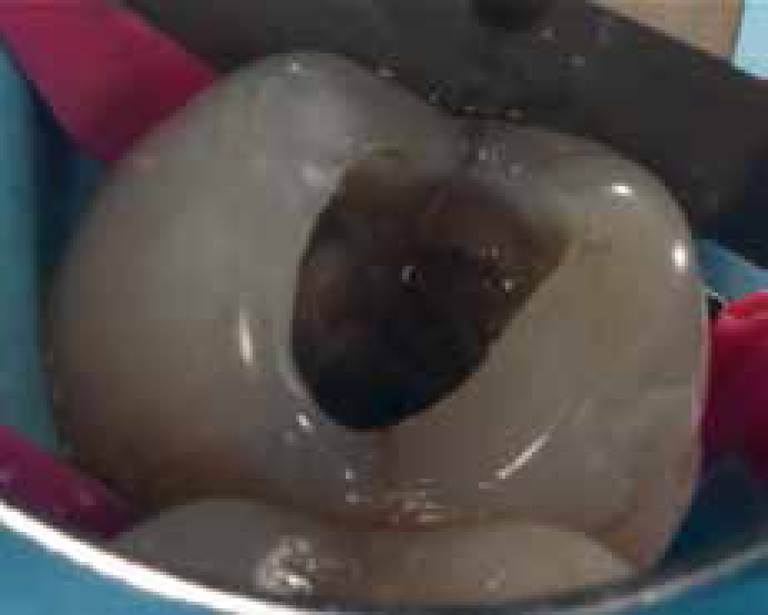
The existing amalgam restoration was removed with a pear-shaped diamond burr (Brasseler, USA) in an electric handpiece with copious water irrigation and microscope-enhanced vision (Ziess Pico, NuView, Glos, UK) (Figure 3). Caries was noted globally throughout the MO cavity; this was removed with a tungsten-carbide rosehead burr. The margins of the cavity were flared and finished with an EMS Cavity Prep Tips SM (Optident, Ilkley, UK) in a piezon scaler unit (EMS Minipiezon, Optident) to reduce the risk of collateral damage to the adjacent canine tooth. The entire tooth was then air abraded with 27-micron alumina (Micro-Etcher, Danville Materials, CA, USA) to remove residual biofilm and aprismatic enamel, therefore enhancing bond strength.
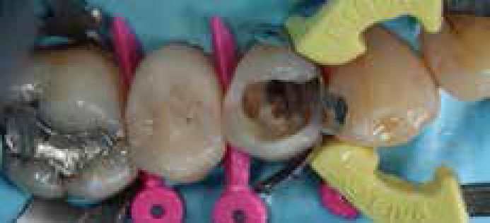
A matrix was placed using the Triodent V3 System and Pink Wavewedge: this was burnished against the adjacent tooth with a cotton pellet in locking tweezers to avoid scarring the soft metal matrix, since scars on the matrix transfer to roughness in the contact area of the final restoration (Figure 4).
The cavity was etched: 15s dentine and 30s enamel with 37% phosphoric acid (Ultra-Etch, Ultradent, UT, USA) washed with air-water spray and dried according to the ‘Wet-Bonding’ Protocol. A 4th Generation Dentine Bonding Agent was applied (Optibond FL) and polymerized for 30s as per manufacturer's instructions (Figure 5).
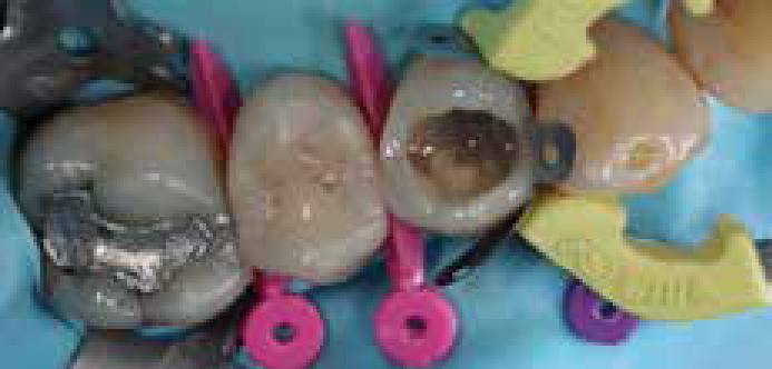
The mesial proximal wall was built with paste resin composite (G-Aenial, GC); shade Adult Enamel (AE). The wall was restored in two separate vertical elements, polymerizing the first for 3s (as per the ‘Pulse-Activation’ protocol), before proceeding to the second, in order to reduce polymerization shrinkage stress. A very thin layer of flowable composite resin (G-Aenial Flo, GC), around 0.5 mm in section was adapted to the cavity floor and photo-activated for 40 s.
A capsulated autocure Resin-Modified Glass Ionomer Cement (Fuji VIII GP, GC) was mixed and injected into the cavity around 1 mm gingival to the amelo-dentinal junction. This was condensed with a microbrush lightly soaked in Optibond FL adhesive and allowed to autopolymerize for 3 min. The bonding agent was then photo-activated for a further 30 s (Figure 6).
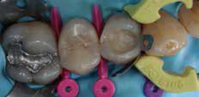
Composite resin (G-Aenial A3 and AE, IL, USA) alongside subtle resin tints (Brown2, Micerium, GDF mbH, Germany) was layered in an incremental cusp-by-cusp fashion polymerizing each cusp increment for 3 s before placing the next. The matrix system was removed and minor marginal finishing carried out with a number 12-scalpel blade (Swann-Morton) on a Bard-Parker Handle (Figure 7).
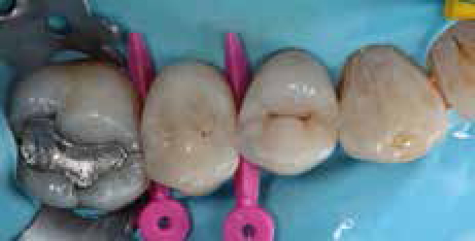
The rubber dam was removed and the occlusion was verified in both static and dynamic movements with articulating paper: no adjustments were required. Contacts were checked with dental tape. KY Jelly (Johnson and Johnson, Maidenhead, Berks) was applied and a final photo-polymerization of 40 seconds carried out to remove the oxygen-inhibited layer. No further polishing was required (Figure 8).
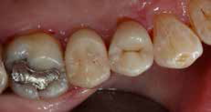
The patient was dismissed. At telephone review some 24 hours later, the patient revealed that his symptoms had completely subsided. At 6-month review the tooth continued to be asymptomatic and there was no deterioration in the restoration or the underlying tooth substrate.
