Article
This paper presents a novel clinical technique of employing an e.max lingual retainer combined with a natural tooth pontic to manage the immediate restoration of a post-extraction space in the lower anterior region. It presents an interesting case report and discusses the use of the e.max lingual retainer compared to other techniques available for managing the immediate restoration of a post-extraction space.
The loss of an anterior tooth can present a challenging clinical situation with regards to the aesthetic, functional and psychosocial concerns for the patient, in addition to the difficulty of restoring such an edentulous space in a timely if not immediate manner.
Case report
An 80-year-old female patient presented complaining of a loose lower front tooth. She explained that the tooth had become progressively loose and, although currently asymptomatic, she had suffered multiple abscesses. Medically, this patient suffered from rheumatoid arthritis and back pain. She had previously been diagnosed with Malignant Peripheral Nerve Sheath Tumour (MPNST) in the post nasal space with extension to the left orbit and frontal lobe. This had been treated by endoscopic craniofacial resection of the tumour and radiotherapy in 2013. She had subsequently undergone a nasal reconstruction procedure.
She found attending appointments difficult, having a long journey and relying on hospital transport. Owing to her back, it was also difficult to sit in the dental chair for long periods. She had been a regular attender to her general dental practitioner, brushed twice a day with a manual toothbrush but admitted that brushing was challenging owing to the effects of rheumatoid arthritis on her hands.
Extra-oral examination was unremarkable except for nasal collapse (Figure 1). She had a young biological age and took great care in her appearance. On speaking and smiling the lower incisors were visible, but the gingival margins did not show.

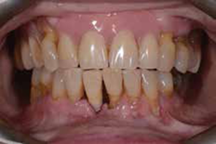
Intra-oral examination (Figure 2) demonstrated normal soft tissue appearance and a good quality and quantity of saliva. She was partially dentate, the teeth present being:
| 3 | 345 |
| 54321 | 12345 |
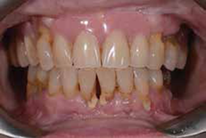
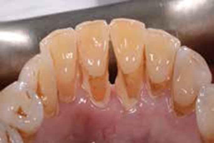
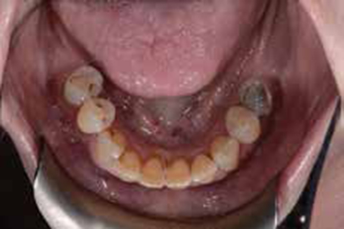
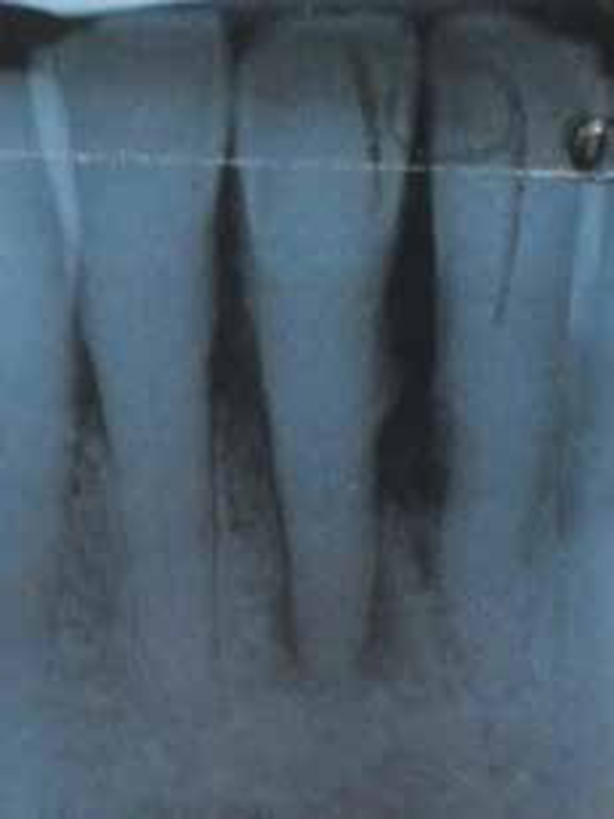
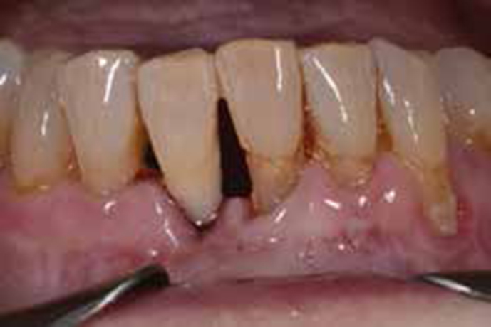
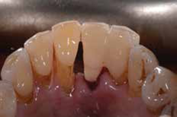
She wore a partial acrylic resin denture in the maxillary arch replacing the incisors and right premolars. There was a reduced mandibular dental arch and obvious supragingival calculus and plaque deposits in each sextant. There was no caries. The LR1 and LL1 were grade 1 mobile, whilst the LR1 was also tender to percussion and had a negative response to sensibility testing with ethyl chloride. The LL1 and LR2 were not tender to percussion, had vital responses to ethyl chloride and were asymptomatic (Figures 3, 4). The LR2 was not mobile. A periapical radiograph (Figure 5) of LR2, LR1 and LL1 demonstrated a vertical pattern of bone loss of 80% around the lower central incisors and 50% on the LR2. Supra- and subgingival calculus was clearly visible interdentally. A periapical radiolucency was apparent on LR1, as was sclerosis of the pulp canal.
Diagnosis
Treatment options for LR1
Accepting the poor prognosis for the LR1 and considering the patient's social information, she decided to have an extraction. She was concerned about appearance and would not accept a space even temporarily, preferring an immediate fixed replacement. One option might have been the use of resin fibre splint from the LR2 to the LL1, either building a tooth in composite resin or using the crown of the LR1 as a natural tooth pontic. The mobility of the LL1 and her difficulty with sitting in the dental chair for a long period made this option less desirable.
Instead, a novel approach was employed, making use of a natural tooth pontic supported by an IPS e.max retainer, extending from the lingual surface of the LR2. This allowed for an aesthetic, fixed, immediate replacement option. This could possibly serve as a long-term treatment modality.
Treatment stages
Following a stabilization stage, including oral hygiene instruction, in particular, use of electric brush and scaling, the treatment was performed as follows:
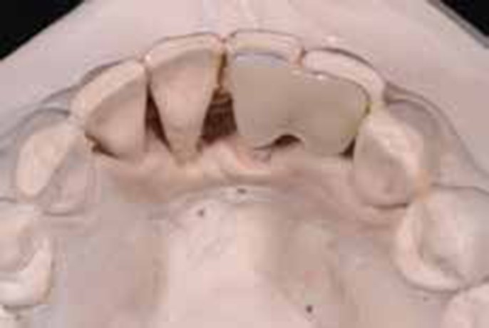
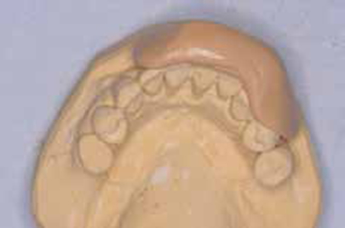
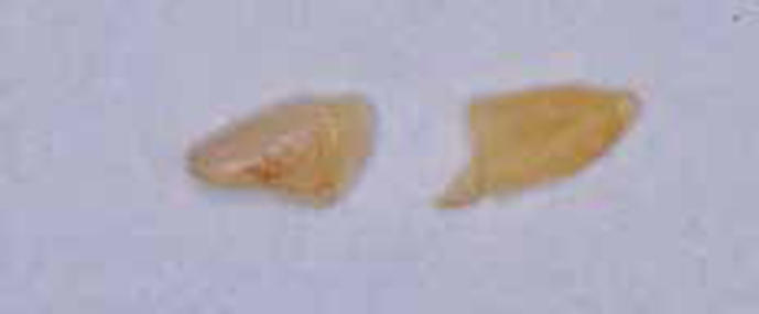
Figures 9, 10 and 11 present postoperative views.
Discussion
Anterior teeth are often lost due to traumatic injury or periodontal disease. The number of solutions for immediate replacement have been described, including the provision of a single tooth on a removable acrylic prosthesis, use of a prefabricated acrylic tooth as a pontic splinted to adjacent teeth, immediate bridgework or an implant.
If not grossly carious or discoloured, an extracted tooth can be used as a natural pontic and splinted to a stable adjacent tooth. The natural pontic provides the characteristics of colour, shape, size and alignment without the need for substantial modifications and avoids the patient having to wear a removable prosthesis. This approach has been described in the dental literature for over 30 years. It enhances the psychological and social acceptability of an extraction and offers a conservative option for tooth replacement.
For this approach to be used, the abutment tooth must be intact or minimally restored and have substantial enamel available for bonding. Candidates for a natural tooth pontic include periodontally involved teeth, teeth with fractured roots and teeth unsuccessfully replanted after avulsion or following failed endodontic procedures. Contra-indications include parafunctional habits, insufficient coronal tooth structure, insufficient occlusal clearance for a splint and an inability to maintain isolation during bonding procedures.
Techniques described include pure composite resin, twist-flex wire, metal or nylon meshwork, fibre reinforced composite resin, a cast metal framework1 and even one technique describing the use of palatal archwire and orthodontic bands.2 Many of the options could be available for use at an emergency appointment.
IPS e.max is an all-ceramic material used in dentistry for inlays, veneers, crowns and bridges.3 Available from Ivoclar Vivadent, the IPS e.max system offers materials for both Press and CAD/CAM applications. Being a lithium disilicate glass ceramic, it has a flexural strength of 400 MPa4 and, owing to its ability to be etched, can form a strong resin–ceramic bond through the use of hydrofluoric acid etching and silane application.5 All-ceramic prostheses can also provide an aesthetic and biocompatibility advantage when compared to metal-ceramic structures.6 It has therefore been used to great effect in anterior fixed prosthesis.7,8
The cost of a laboratory constructed ceramic retainer compared to a metal retainer or a direct technique should be considered and may present a barrier to its use. In addition, the evidence for resin-bonded bridges supports the use of metal retainers for resin bonding over all-ceramic or fibre-reinforced resins in terms of long-term survival.9,10 A literature review estimated the annual failure rates per year to be 4.6% for metal-framed, 4.1% for fibre-reinforced and 11.7% for all-ceramic resin-bonded bridges.11 The most frequent complications include debonding of metal-framed resin-bonded bridges, delamination of the composite veneering material in fibre-reinforced bridges, and fracture of the framework of all-ceramic bridges. In this case, the NTP was opposed by a removable acrylic resin tissue-supported denture which would reduce occlusal load on the bridge compared to a natural dentition or fixed restoration.
Conclusion
The use of a natural tooth pontic in this 80-year-old female patient allowed an immediate, aesthetic and functional result which permitted good oral hygiene and prevented the need for a removable partial denture. Considering the patient's age and evidence relating to longevity of e.max bridgework, this restoration will be monitored as a long-term treatment modality.
