Article
Response to glandular odontogenic cyst – a case report
We read with interest Manouchehri and Ali's case report of a glandular odontogenic cyst affecting the LR67 region.1 We too present a recent case of a glandular odontogenic cyst presenting in an otherwise healthy 61-year-old male.
The patient was referred to the Department of Oral and Maxillofacial Surgery at Royal London Hospital by his GDP regarding the recurrence of a cyst which was enucleated in 2010, along with removal of the LR8. The histopathology reported at the time was consistent with a dentigerous cyst associated with an unerupted lower right third molar. The patient was regularly reviewed, with satisfactory healing and normal neurosensory function leading to his eventual discharge.
In this new episode, the patient complained of occasional pain from the area where the cyst was previously enucleated. There were no other complaints. Clinical examination revealed no neurosensory deficit and an unremarkable extra-oral appearance. There was, however, a palpable bony expansion felt in the right body of the mandible. The LR7 was non-mobile and buccal expansion was noted in the right posterior mandible. An egg-shell type crackling was also apparent lingually. The overlying mucosa was otherwise normal.
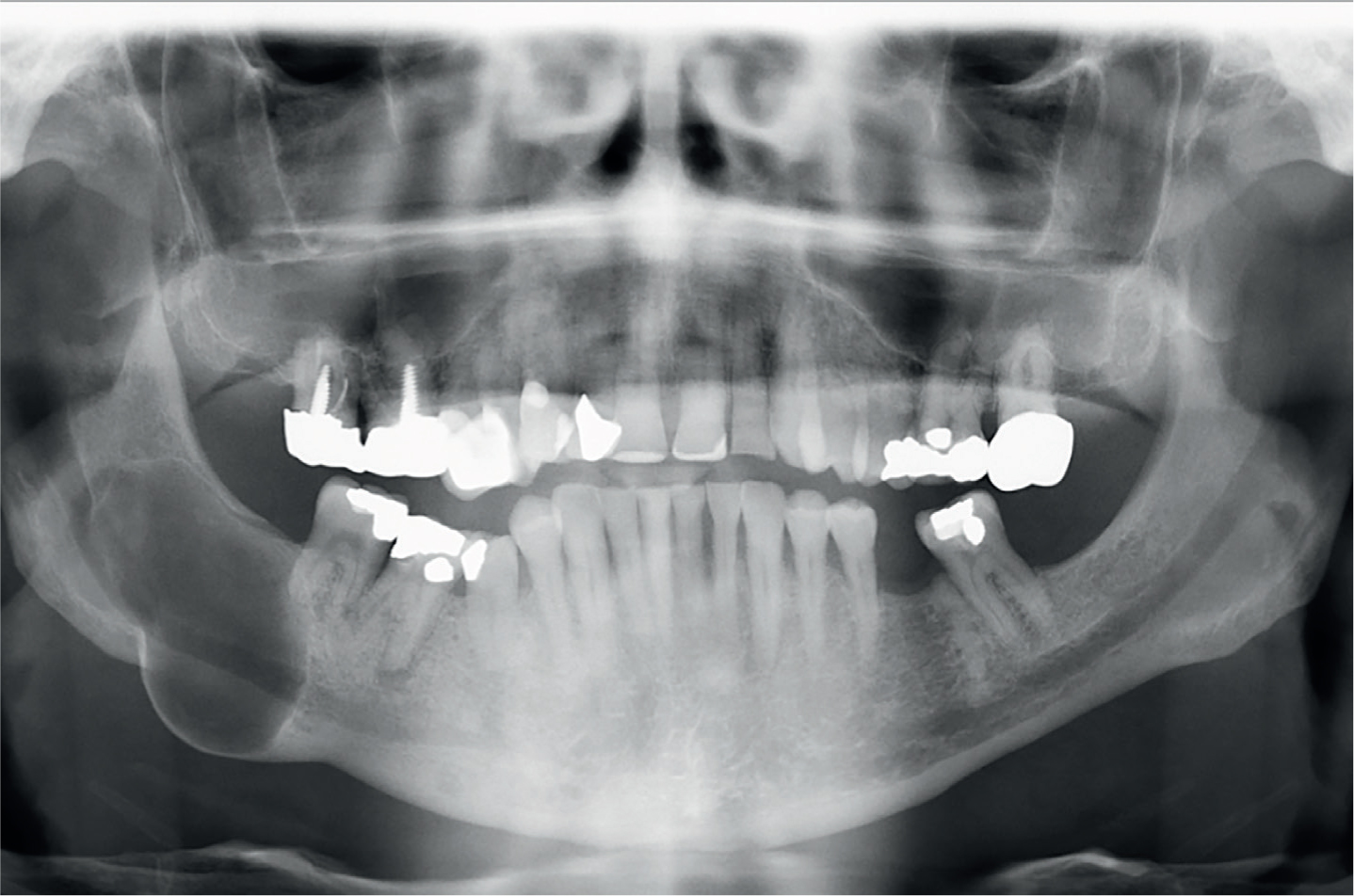
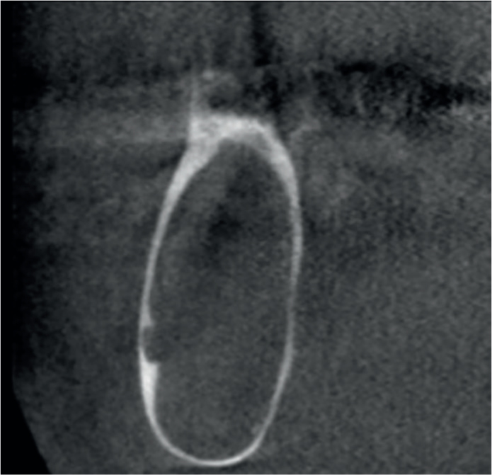
Radiographic examination revealed a unilocular, corticated, radiolucent lesion extending from the ascending ramus to the furcation of the LR7 (Figure 1). There was significant thinning of the lower border of the mandible. Coronal views on CBCT imaging displayed the extent of bone resorption with loss of cortication on the inferior dental canal, which was buccally placed (Figure 2).
The cyst was enucleated uneventfully under general anaesthetic, followed by a strict post-operative regimen of a soft-diet for 8 weeks. At early follow-up, the patient reported no issues following his procedure and no neurosensory deficit.
The histopathology demonstrated a cystic cavity lined by non-keratinizing, stratified, squamous epithelium. The presence of mucous metaplasia and intra-epithelial ductal structures with plaques in the epithelial lining raised the likelihood of this specimen representing a glandular odontogenic cyst.
As discussed, glandular odontogenic cysts are reported often to be misdiagnosed due to their non-specific features.1 At initial presentation, the presence of an unerupted LR8 may have misled/influenced the final diagnosis into being a dentigerous cyst.