Article
Unilateral trifid mental nerve and foramen: a case report
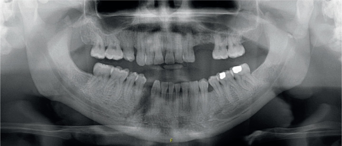
A 29-year-old African male patient presented with a fractured right parasymphysis and left condylar base following an altercation while under the influence (Figure 1). On examination the patient had a deranged occlusion with mobility of the anterior mandibular segment. He had an apparent skeletal base Class 3 and an anterior open-bite, yet incisal edge wear facets indicated an edge-to-edge occlusion prior to the incident. Plain film imaging demonstrated significantly reduced height of the left condyle and displacement of the right parasymphyseal fracture (Figure 1). The patient was admitted and consented for open reduction and fixation of both fracture sites.

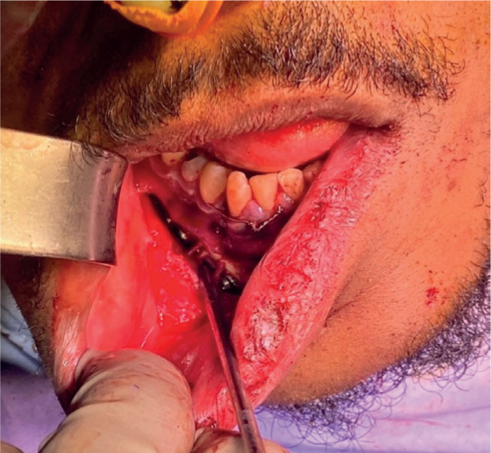
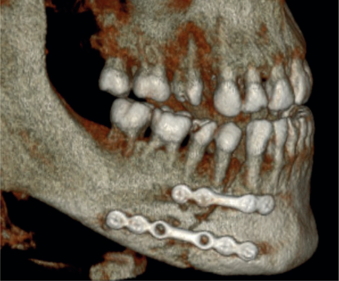
Interestingly, during access to the right parasymphyseal fracture, three separate mental nerves and foramina were discovered unilaterally. It was initially thought that there was only a second foramen and associated nerve; however, further exploration of the surgical field revealed a third foramen and nerve. All three foramina and mental nerves were anatomically distinct from each other and had comparative diameters (Figure 2). It was unclear which was anatomically dominant, and which were accessory foramen. All three nerves were preserved during reduction and plating with some post-operative paraesthesia to the lower lip observed clinically. A CBCT was acquired post operatively due to possible fracture displacement. A consultant radiologist reviewed the CBCT independently, confirming the finding of a unilateral triplicate nerve with a single mental foramen contralaterally (Figure 3).


Variation in mental nerve anatomy is widely reported, ranging from multiple mental nerves and foramen to complete absence.1 Detection of such variations is increasing alongside imaging availability, with retrospective studies based on analysis of CBCT/CT records2–5 reflecting the potential limitations of pre-operative plain film radiographs.
Although some clinicians recommend pre-operative CBCT for surgical procedures,3 this must be justified depending on the planned procedure, in line with national and local radiography guidelines.6 As such, an awareness of significantly variable anatomy promotes increased caution for the practitioner and, perhaps, the additional need to inform and consent a patient of the innate operative risk accompanying individual anatomy.
We hope to illustrate the potential for significant anatomical variation among patients with key points from this letter applying to all dental professionals:
Ultimately, a cautious, considered surgical approach to prevent avoidable morbidities should always be employed.
