References
A case of extensive oral kaposi's sarcoma in a patient with undiagnosed HIV infection
From Volume 45, Issue 4, April 2018 | Pages 360-362
Article

Kaposi's sarcoma is a low grade, soft tissue tumour of vascular endothelial origin. In 71% of cases, it presents with cutaneous and visceral involvement and most commonly arises amongst the male population.1,2 In 1994, Chang and colleagues identified a new herpesvirus (HHV-8) which was at the time linked with over 95% of Kaposi's sarcoma (KS) lesions identified.3 Although infection with the HHV-8 virus is now considered necessary for development of the tumour, it is not sufficient alone, and a variety of other co-factors appear also to be involved.4
Infection with HIV is one such co-factor and the incidence of KS is approximately 1 in 20 amongst individuals infected with HIV.5 In around 22% of cases, KS manifests with oral involvement as its first presentation.2
Here the case of a patient who presented with oral, cutaneous and visceral lesions associated with KS is discussed on a background of undiagnosed HIV infection.
Case report
A 25-year-old male presented to the Emergency Department of Royal Gwent Hospital with a 6 day history of dyspnoea, severe headaches and a self-terminating episode of supraventricular tachycardia (SVT). He also had multiple oral and cutaneous lesions which had been present for several months. The patient's medical history comprised asthma, atopic dermatitis and hayfever. He had one episode of SVT 8 months prior to this admission and had a 10 pack year smoking history.
On examination, there were multiple, well-defined, bluish-red lesions on both the left upper and lower eyelids (Figure 1). These were non-tender on palpation, soft and non-blanching. There were several similar lesions on the sclera of both eyes (Figure 2).


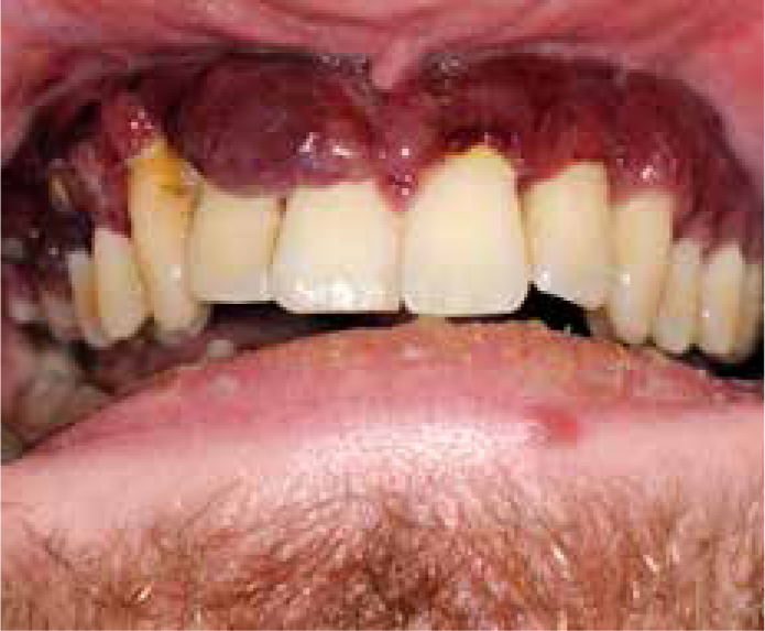
Intra-oral examination revealed a diffuse bluish-red swelling involving almost the entire hard palate with several nodular type lesions present (Figure 3). On palpation, these were firm with no tenderness or discharge noted. Corresponding lesions were present on both the upper and lower gingiva (Figure 4), with no associated mobility of teeth.


A new diagnosis of HIV was made by the medical team and a lung biopsy confirmed the clinically suspected KS which had visceral, mucocutaneous and pulmonary involvement. The orofacial lesions clinically supported the diagnosis of Kaposi's sarcoma and it was felt that a further intra-oral biopsy was unnecessary to confirm diagnosis.
Prior to this acute presentation, the patient had been referred to hospital on two separate occasions regarding both the oral and associated skin lesions. Having attended for assessment in the maxillofacial unit six months earlier, the patient had failed to attend for any further investigation and was lost to follow-up.
He was commenced on palliative chemotherapy with liposomal doxorubicin and highly active antiretroviral therapy (HAART) was initiated, comprising ritonavir and emtricitabine/tenofovir. The sarcoma lesions failed to regress with treatment and the patient died one month following this admission to hospital.
Discussion
Kaposi's sarcoma often presents as a slowly progressive tumour, although in some cases it can present in a more aggressive manner.5 Traditionally, there are four main classifications of KS including:
HAART represents a combination of drugs used to suppress the viral load of HIV. Its introduction has revolutionized HIV treatment and resulted in a steep decline in the prevalence of oral lesions associated with HIV, including hairy leukoplakia and herpes simplex labialis, amongst others.7 Although this has also been the case for epidemic Kaposi's sarcoma, it remains to be the second most common tumour amongst HIV-infected patients worldwide.5,8
In 2012, the prevalence of HIV infection was estimated to be approximately 35,300,000 worldwide.8 It is of particular importance therefore that clinicians, especially oral health providers, are able to recognize clinical presentations of HIV-associated disease.
The overall prognosis of KS is dependent on a number of factors, including the level of immunosuppression, viral load and the overall extent of the condition.8,9 In order to recognize the lesion, we first need to be aware of its clinical presentation. With KS, there are a number of stages that can present. Because of this it can mimic several other benign lesions, potentially leading to a mistaken diagnosis.10,11
There are three distinct clinical stages of KS including:
The patch stage is characterized by pink, purple or red macules on the oral mucosa and can resemble lesions such as thermal trauma, haematoma or atrophic candidiasis.12 Often at this stage the histological features of KS are not yet evident.12The lesion develops to the plaque stage where it increases in size becoming raised and violaceous in appearance.13,14
Finally, as the tumour continues to grow, it can form nodular exophytic lesions.13 At both plaque and nodular stages, it can mimic pyogenic granuloma, haemangioma and angiosarcoma, amongst others.12 Occasionally, these lesions can be surrounded by a yellow-coloured mucosa and, in later stages, become ulcerated or infected, sometimes causing resorption of surrounding bone.15 Pain and tenderness are not usually a feature of early disease but do become more common in the later stages.15
Although the tumour may develop on any mucosal site, it most commonly presents on the hard palate, soft palate and gingivae.2,9 This tumour can represent a significant burden on patients in terms of both quality of life and aesthetics.2 Treatment focuses on halting growth and in some cases causing complete regression of the disease, as well as management of the underlying cause. Due to the multifocal nature of the disease, surgical intervention is often limited, with chemotherapy, radiotherapy and HAART forming commonly used treatment modalities.2,16
Conclusion
Although in recent decades there has been an overall decline in epidemic Kaposi's sarcoma, when tumours do arise the high proportion presenting initially on the oral mucosa makes awareness of the tumour by dental professionals particularly important.5,8 Their frequent provision of oral examination combined with knowledge of the tumour and its clinical features may make referral and diagnosis of disease more efficient. Additionally, this case highlights the importance of universal precautions with regards to infection control, as patients may attend the primary dental care setting with subclinical viral infection, as was apparent for many years with the patient described. While the clinical trajectory of KS is multifactorial, it remains true that in all cases early diagnosis is key to improving overall patient prognosis.
