The orthodontic-surgical management of a patient with gorlin syndrome: a case report Madeleine Storey Susi Caldwell Simon Watkinson Manu Patel Dental Update 2025 46:11, 1056-1061.
Authors
MadeleineStorey
BDS(Hons), MFDS, MRes, MOrth, Post CCST Registrar in Orthodontics, Manchester Foundation Trust
This case report describes the management of a patient following the incidental finding of multiple odontogenic keratocysts on an orthopantomogram by the patient's general dental practitioner (GDP). The cysts were extensive and had caused considerable displacement of the unerupted permanent teeth. Following marsupialisation, the teeth were aligned orthodontically. This article describes the features of odontogenic keratocysts, the associated Gorlin syndrome, and the management options available. The importance of close collaboration between the Oral and Maxillofacial Surgical and Orthodontic teams is highlighted.
CPD/Clinical Relevance: Odontogenic keratocysts are benign but locally aggressive jaw cysts. They occur most commonly as solitary lesions in the jaws of healthy individuals, but may also be a feature of Gorlin syndrome. In young patients there is potential for severely displaced teeth to improve their position spontaneously and erupt.
Article
Madeleine Storey
Odontogenic keratocysts (OKCs) were first described in the literature by Philipsen in 1956.1 They are thought to arise from the remnants of the dental lamina which persists after the completion of odontogenesis.2 OKCs are benign but locally aggressive. They occur most commonly as solitary lesions in the jaws of healthy individuals and show a high incidence of recurrence if not adequately removed.
Epidemiological data vary considerably: OKCs account for between 2% and 11% of all jaw cysts and can occur at any age. They are more common in males than females with a male:female ratio of approximately 2:1. However, this is closer to unity in Caucasian populations and greater in Afro-Caribbean patients.3
Clinical presentation
Odontogenic keratocysts are usually asymptomatic and are, therefore, commonly diagnosed as incidental findings on dental radiographs, often whilst investigating the cause and location of unerupted teeth. When they do cause symptoms, these can be in the form of pain, swelling and discharge, often as a result of secondary infection. The majority (over 70%) occur in the mandible with approximately half of these occurring at the angle of the mandible.3
Unlike most other jaw cysts which expand by osmotic pressure, OKCs expand due to increased epithelial turnover, therefore bony expansion is not a common finding.
Radiographically, they may present as uni- or multi-locular radiolucencies with a differential diagnosis of radicular, residual or dentigerous cysts.
Management
The traditional method for the treatment of most OKCs is surgical enucleation. Marsupialisation is preferred for larger cysts, often followed by enucleation. Adjunctive surgical treatments, such as removal of the peripheral bone (ostectomy) or resection of the cyst, together with surrounding bone (en-bloc resection), have also been proposed to minimize the chance of recurrence.4–6 Cryotherapy with liquid nitrogen,7 and the use of Carnoy's fixative solution8 in the cyst cavity following enucleation, have also been suggested in an attempt to address any remaining cystic tissue and minimize recurrence.
In 2015, a Cochrane systematic review investigated treatment of OKCs. The authors stated that, currently, no conclusions can be made about the effectiveness of the interventions available for the management of OKCs.9
Gorlin syndrome
Gorlin syndrome is an autosomal dominant condition which occurs with equal frequency in both sexes. It has both a familial and a sporadic incidence, with 20–40% of cases occurring as de novo mutations, and a reported prevalence of 1 in 56,000 to 164,000.10 Gorlin syndrome has been shown to be caused by loss or mutation of the PTCH1 (patched) gene found on chromosome 9q.11
Gorlin syndrome is a multi-system condition that can cause an altered facial appearance due to changes in the cranial base, and a predisposition for basal cell carcinoma (BCC) in atypical sites not chronically exposed to sunlight. OKCs in Gorlin syndrome appear in a younger age group, typically during the first decade of life, more commonly in the mandible than in the maxilla, and may be single or multiple. They develop in 65% to 75% of patients with Gorlin syndrome and have a high risk of recurrence.12 Diagnosis of Gorlin syndrome is made by having two major criteria or one major and two minor criteria (Table 1).
Major criteria
Minor criteria
Occurrence of two or more BCCs in patients younger than 20 years
Radiographic abnormalities; bridging of the sella turcica, vertebral anomalies, malformations of hands and feet
Rib deformities (fused or bifid)
Ovarian and cardiac fibromas
First-degree relatives with NBCCS
Medulloblastoma
Patients with Gorlin syndrome need educating about the syndrome. Exposure to UV light should be reduced to minimize the risk of developing BCCs. Genetic counselling is advised since one parent with Gorlin syndrome results in a 50% probability that his/her child will be affected. Treatment is usually supportive to minimize symptoms rather than to cure the condition. Enucleation of OKCs can help but new lesions, infections and jaw deformities may result. The severity of BCCs determines the prognosis for most patients. BCCs rarely cause gross disfigurement, disability or death, but can metastasize.
Case report
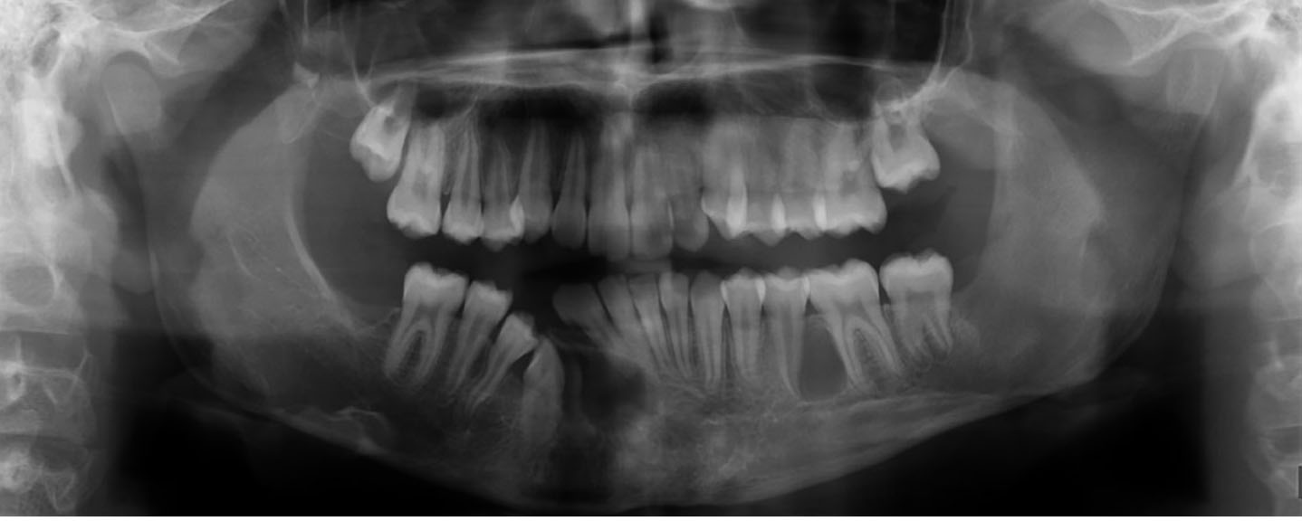
A 9-year-old girl was referred urgently to the Oral and Maxillofacial Surgery Department after her GDP noticed a large radiolucent area on an orthopantomogram (OPG) and suspected neoplasia. Clinically, the crowns of lower anterior teeth were tilted to the right and reactive bony expansion was palpable buccally in the right premolar region. Radiographically, three large unilocular radiolucencies with well demarcated margins were detected in the body of the mandible, displacing the unerupted lower right permanent canine and premolars and both lower second molars (Figure 1a). A provisional diagnosis of OKCs was made.
Figure 1. (a) Initial orthopantomogram showing large unilocular radiolucencies displacing the unerupted lower right permanent canine and premolars and both lower second molars.
Due to the significant size of these cystic areas and the associated risk of fracture of the mandible, devitalization, or loss of the displaced teeth, the decision was made to marsupialise rather than enucleate the cysts.
Peri-operatively, the lower right second molar (LR7) was found to be floating free in the cyst cavity and deemed to be of poor long-term prognosis, therefore it was removed. Tissue samples confirmed the provisional clinical diagnosis of OKCs. Post-operatively, the patient was referred to the Orthodontic Department for an opinion on the management of the unerupted displaced teeth.
The patient presented in the mixed dentition with a Class I incisor relationship on a Skeletal I base with average vertical proportions. The upper arch was well aligned with both upper permanent canines palpable buccally. The lower anterior teeth were moderately crowded with the lower dental centreline displaced to the right.
The three-month post-operative OPG showed some improvement in the position of the unerupted, displaced permanent teeth following marsupialisation of the cysts. The root formation of these teeth was less than one-fifth that of a normal root, suggesting eruptive potential (Figure 1b). Combined with the extent of bone loss due to the previous presence of cysts, the decision was made to monitor their position and further development. Serial radiographs taken post-operatively confirmed both bony infilling and spontaneous improvement in the position of the unerupted teeth (Figure 1c–f). The patient was warned that there was no guarantee of eruption but, as she was young, there was significant eruptive potential.
Figure 1. (b) Three months post marsupialisation.Figure 1. (c) Twelve months post marsupialisation.Figure 1. (d) 2½ years post marsupialisation.Figure 1. (e) 3 years post marsupialisation.Figure 1. (f) End of orthodontic treatment: note the positions of the developing upper third molars, displaced by the previous presence of odontogenic keratocysts.
The patient was monitored closely by both the Oral and Maxillofacial Surgery and Orthodontic Departments. Further surgery was necessary to remove cyst remnants and recurrences, whilst a soft tissue exposure of the LL7 was performed to encourage its eruption into a more favourable position. Due the observed high recurrence rate of the OKCs, the patient was referred for genetic testing to investigate the possibility of Gorlin syndrome. Diagnosis was confirmed based on the presence of multiple, recurrent OKCs, palmar and plantar pits, macrocephaly, and calcification of the falx cerebri.
Three years after diagnosis, the previously displaced teeth had erupted sufficiently to commence orthodontic treatment (Figure 2). Following an initial period of aligning, levelling, space creation and centreline correction, an attachment was bonded to the partially-erupted, previously grossly displaced lower right canine (LR3) to which an inter-arch elastic was placed to encourage eruption (Figure 3). After the canine had erupted sufficiently, residual spaces were closed with both intra- and inter-arch elastics, and the occlusion was detailed and finished. The fixed appliances were removed after 28 months of active treatment and upper and lower vacuum-formed retainers were fitted to wear indefinitely on a nights-only basis (Figure 4). The patient continues to be monitored clinically and radiographically, where appropriate, by the Oral and Maxillofacial Surgery team for identification and subsequent management of cyst recurrence.
Figure 2. (a–e) Start orthodontic records.Figure 3. Inter-arch elastic to erupt the lower right canine mechanically.Figure 4. (a–e) End of orthodontic treatment.
Discussion
This case report describes the inter-disciplinary management of teeth displaced by the presence of cysts. When new patients are seen, it is important to account for all teeth not clinically evident in the mouth. Teeth missing from the line of the arch may be developmentally absent or significantly displaced. Good quality radiographs are essential to assess tooth presence, position, and to identify any associated or causative pathology such as cysts. Bitewing and periapical radiographs may fail to demonstrate the true extent of cystic lesions, therefore an OPG or CBCT is likely to allow better assessment.13 Following diagnosis and initial surgical management, radiographic review is important to monitor subsequent tooth eruption, cyst recurrence, and bony infilling.
In paediatric patients, marsupialisation should be the treatment of choice if developing teeth are to be preserved. Marsupialisation consists of fenestrating the outer wall of the cyst into the oral cavity, thus relieving intracystic pressure. Decompression allows the cyst cavity to reduce slowly in size, enabling the surrounding bone to regenerate around the impacted tooth, facilitating the potential for eruption into the dental arch.13 Subsequent enucleation of the cyst lining and histopathologic examination is necessary for definitive diagnosis and to rule out neoplastic transformation.14
Odontogenic keratocysts have a tendency to recur after treatment with recurrence rates ranging from 3% to 60%.3 Many theories have been proposed to account for their high level of recurrence. The cyst lining is delicate and remnants can be left behind after surgical removal, or satellite cysts (from odontogenic epithelial remnants), or daughter cysts (from out-pouchings of the main cyst lining) may develop into new cysts after removal.15
Odontogenic keratocysts in patients with Gorlin syndrome present a different problem from those found in isolation, as these patients have an inherited tendency to develop new cysts, in contrast to local recurrence alone. As such, a conservative surgical approach is advocated; specifically, initial marsupialisation with subsequent enucleation of cysts associated with impacted and unerupted permanent teeth.16
Patients should be closely monitored by the Oral and Maxillofacial Surgery team well into their adult lives. Patient-reported signs and symptoms or clinical evidence of swelling or jaw deformity may be indicative of recurrence and would warrant further radiographic investigation. There are currently no evidence-based guidelines for the duration of long-term follow-up or the recommended frequency of radiographic assessment, so each case should be judged and managed individually.
The option to preserve teeth associated with cysts should be considered as even significantly displaced teeth often have the ability to erupt towards the line of the arch following the removal of an obstruction. This should be considered when discussing treatment options during the consent process. Early communication with an orthodontist would therefore be recommended.
It has been reported that the eruption speed of an impacted tooth associated with a cyst is faster three months after marsupialisation than at other times, regardless of the degree of root formation or type of cyst. Orthodontic traction of an impacted tooth with an immature root may cause root resorption, root deformation, or pulpitis. As such, traction was not applied until root maturation was observed. In this case, the position of the impacted teeth improved remarkably following marsupialisation and they successfully erupted. However, traction may need to be applied in older patients where the eruptive potential has been lost and root formation is complete.
Conclusions
Odontogenic keratocysts are benign but locally aggressive jaw cysts, most commonly occurring as solitary lesions in the jaws of healthy individuals, but may also be a feature of Gorlin syndrome. These cysts tend to grow rapidly, are prone to recurrence, and may cause displacement of developing teeth. Management is often interdisciplinary to treat both the cysts and any associated or affected unerupted teeth. In young patients there is potential for severely displaced teeth to improve their position spontaneously and erupt.