Lou Q, Wang X, Jiang L Subjective and objective evaluation of speech in adult patients with unrepaired cleft palate. J Craniofac Surg. 2022; 33:e528-e532 https://doi.org/10.1097/SCS.0000000000008567
Pegoraro-Krook MI, Rosa RR, Aferri HC, Andrade LKF, Dutka JCR Pharyngeal bulb prosthesis and speech outcome in patients with cleft palate. Braz J Otorhinolaryngol. 2022; 88:187-193 https://doi.org/10.1016/j.bjorl.2020.05.028
Yenisey M, Cengiz S, Sarikaya I Prosthetic treatment of congenital hard and soft palate defects. Cleft Palate Craniofac J. 2012; 49:618-621 https://doi.org/10.1597/10-016
Keyf F, Sahin N, Aslan Y Alternative impression technique for a speech-aid prosthesis. Cleft Palate Craniofac J. 2003; 40:566-568
Kahlon SS, Kahlon M, Gupta S, Dhingra PS The soft palate friendly speech bulb for velopharyngeal insufficiency. J Clin Diagn Res. 2016; 10
Speech bulb prostheses serving to restore missing soft palate are becoming less common in modern practice. Methods and techniques including material choice have changed, with the majority of reference literature now appearing outdated for newly qualified practitioners. This article illustrates how an archaic prosthodontic complication was managed using techniques based on traditional methods.
CPD/Clinical Relevance: A current workflow for an infrequent, archaic prosthodontic challenge is presented.
Article
The presentation of patients with large, unrepaired maxillary defects owing to cleft lip/palate is rare in contemporaneous practice given the more recent advances in surgical techniques and increased availability of reconstructive surgery.1
Modern management of cleft palate often involves surgical management with or without wearing an appliance pre-surgery, with work-up and treatment often starting from birth. In cases where surgical repair of the defect is not possible, hypernasality of speech, masticatory inefficiency and nasal regurgitation manifest.2
In such cases, missing soft and/or hard palate results in palatopharyngeal inadequacy, which often requires obturation by prosthetic means.3
The case presented below illustrates a contemporaneous workflow of a previously common prosthodontic presentation.
Case report
A 95-year-old male patient was referred to the Department of Restorative Dentistry at Newcastle Dental Hospital for a consultation regarding subsatisfactory maxillary complete and mandibular partial dentures. The patient was born with a Veau class IV cleft palate, which was unrepaired (Table 1).4
Class I
Isolated cleft within soft palate only
Class II
Hard and soft palate cleft only
Class III
Unilateral cleft lip and palate
Class IV
Bilateral cleft lip and palate
The patient's main complaint pertained to an unretentive upper complete denture which had an acrylic projection posteriorly to obturate the defect. This was made many years ago. As a result, he was using the fitting surface of the denture to chew with against his hard palate, which resulted in frequent nasal blockage and aspiration. Speech was also unintelligible. Cosmetic concerns were also cited, but the principle complaint pertained to function.
Medically, the patient was diagnosed with a history of transient ischaemic attack, chronic asthma, chronic obstructive pulmonary disease and atrial fibrillation.
On examination, extra-oral findings were unremarkable.
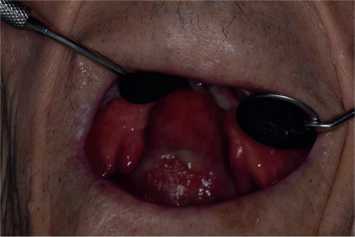
Intra-orally, an edentulous maxillary arch with a maxillary defect that breached the soft and hard palate was noted, with uvula deviation to the right-hand side. The maxillary arch ridge was firm and of reasonable form (Figure 1). The mandibular arch was part dentate with LR2 root remnant and LR3 present which did not exhibit clinical signs or symptoms of endodontic pathology. LR3 was over-erupted. The mandibular arch ridge was of reasonable form, but resorbed. The maxillary complete denture was unretentive.
Figure 1. Unrepaired bilateral cleft involving soft and hard palate absence.
Challenges noted involved managing the patient's medical history, the lone-standing, over-erupted LR3 that could complicate registration, and how to undertake an adequate impression. The aims of treatment were to achieve sound obturation of the defect to prevent nasal regurgitation while not impinging on nasal breathing, and for the denture component of the prosthesis to provide cosmetic improvement.
Ideal images were difficult to take given the patient's inability to be laid back owing to his medical condition. The authors, however, feel that the images suffice to illustrate the general appearance and profile of the defect.
Previous techniques in the literature
Several method workflows exist as to how to manage such a defect, which is impacted by the dentate status of the maxillary arch. In this edentate case, the workflow may well have been simpler than in a dentate case.
A primary impression is often undertaken with a high-tear resistance alginate, which serves as a sound mucostatic impression technique, albeit with the risk of herniating and locking into defects.5
The major impression follows, often involving low temperature fusing compound and wash impression, with the primary aim being to record the maxillary arch form to facilitate registration block fabrication. It was often advocated that this is made with an acrylic base, with a projection posteriorly into the defect. Registration is then undertaken.
Denture try-in, where the denture itself is assessed, is then completed. At this stage, impression for the bulb is undertaken. This traditionally involved reduction of the posterior projection such that the projection just touches the posterior pharyngeal wall, followed by addition of impression wax or compound to the projection which is moulded by simulating functional movements. The aim of additions was to achieve a sound seal of the defect without limiting nasal breathing.
This is processed into acrylic and delivered, then reviewed and subsequent adjustments made if necessary.6
Principles of the technique described were applied to this case, with main changes pertaining to material choice and what was available for our use. The staging workflow was also modified, given our wish to provide a prosthesis as soon as possible given the patient's deteriorating medical status.
Workflow
The primary impression was undertaken using red, high-temperature, fusing cake compound (Kerr, Uxbridge, UK) owing to our wish to achieve a muco-compressive impression. Care was taken to extend the compound into the defect to touch and gently push against the posterior pharyngeal wall. Mandibular impression was undertaken using the compound within edentate areas followed by alginate (Alginoplast, Kulzer, Hanau, Germany) wash.
Special trays were requested. The maxillary tray was specified to be made with a wire loop posteriorly, and the mandibular tray was to be spaced for alginate impression.
The secondary impression of the maxillary arch involved peripheral low-fusing compound impression (Iso Functional Stick Red, GC Corporation, Tokyo, Japan), a silicone putty (VPS Hydroputty, Henry Schein, Melville, USA) addition to the posterior projection to engage into the wire loop for the primary bulb impression, followed by medium-bodied silicone (Aquasil Ultra+ Medium Regular Set, Dentsply Sirona, Charlotte, USA) wash impression. This resulted in a registration block with an acrylic base, and a postero-superior projection intended to gain further obturation of the defect.
Registration was then undertaken. A secondary impression of the bulb was undertaken on the registration block once registration had been completed using low-fusing compound (Iso Functional Stick Red, GC Corporation, Tokyo, Japan), and acrylic (tissue conditioner) reline material (Coe-Comfort, GC America, Alsip, USA) using functional movements.
The patient was instructed to drink, move his head in every direction and talk, particularly to make plosive sounds to mould the posterior extension of the acrylic projection. Nasal breathing was also checked. The aim of this was to mould the borders of the bulb to allow for passive nasal breathing, sound speech and to minimise nasal regurgitation on drinking.
At both primary and secondary bulb impression appointments, the posterior acrylic projection was checked to confirm there was no impingement on the pharyngeal wall, and this was modified if this was the case.
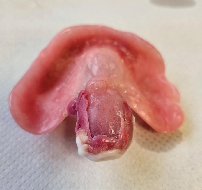
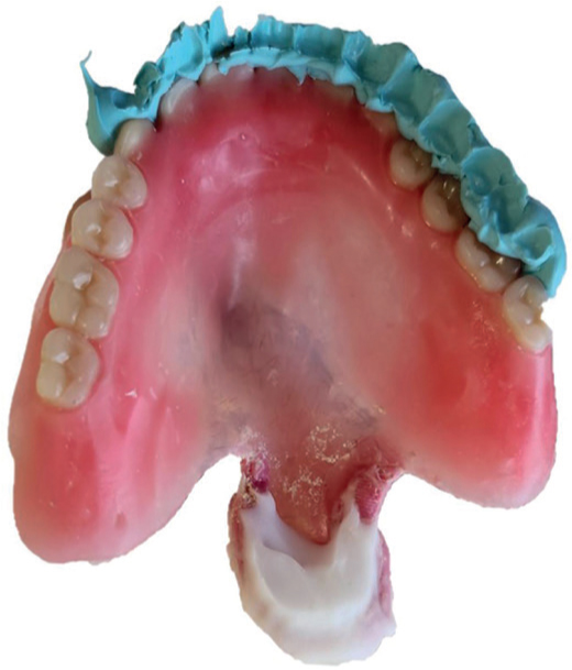
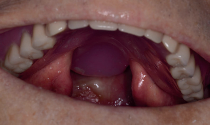
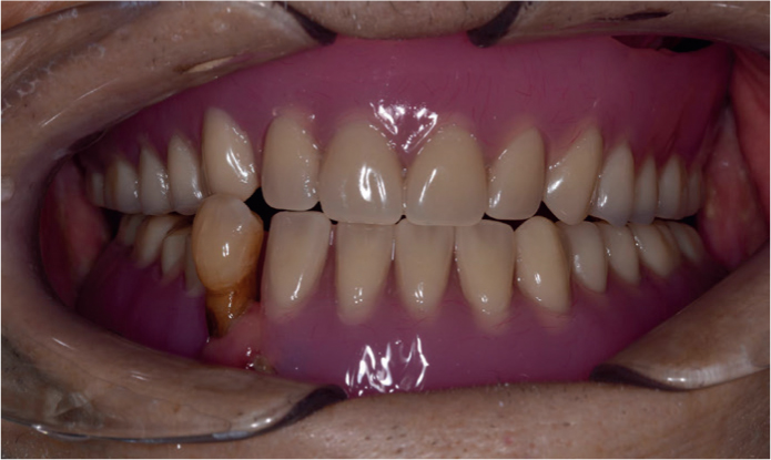
Once a satisfactory bulb impression was achieved (Figure 2), try-in stages (Figure 3) and insert stages were completed, which yielded the final outcome (Figures 4 and 5).
Figure 2. Bulb impression undertaken with compound and tissue conditioner.Figure 3. Final try-in appearance with check record and final bulb impression visible.Figure 4. View of denture in situ with bulb projecting towards posterior wall of pharynx.Figure 5. Anterior view of dentures in situ.
Discussion
Tissue conditioner materials are often useful to trial how to extend and modify a bung, as it can be added and removed easily within a relatively short working time. Indeed, some practitioners advocate the use of tissue conditioner alone without supplementary compound, as was done in this case.7
With regards to staging the prostheses steps, practitioners may advocate treating each component of the prosthesis in a step-wise manner, namely taking the denture to trial stage and addressing the bulb only after satisfactory denture try-in has been achieved.
From a technical perspective, the master cast would require frequent review and sectioning to accommodate for the changes in the dimensions of the bulb.
Conclusion
The above case was successful given the regular review and modification of the bulb from its initial impression. Speech still had a very small element of hypernasality, but this was markedly reduced and speech was intelligible. The patient also did not report nasal regurgitation and was able to breathe through the nose while wearing the prosthesis. He was also satisfied with the cosmetic outcome.
Previous literature outlining stages of provision of such prostheses may appear outdated, particularly given the advent of alternative materials; however, the aims and prosthodontic principles in fabricating a prosthesis remain the same.
The authors recommend using the method and types of materials detailed above to manage this traditionally presenting but rare prosthodontic challenge.