References

Rudy HL, Wake N, Yee J Three-dimensional facial scanning at the fingertips of patients and surgeons: accuracy and precision testing of iPhone X three-dimensional scanner. Plast Reconstr Surg. 2020; 146:1407-1417 https://doi.org/10.1097/PRS.0000000000007387
Koban KC, Leitsch S, Holzbach T 3D Bilderfassung und Analyse in der Plastischen Chirurgie mit Smartphone und Tablet: eine Alternative zu professionellen Systemen? [3D-imaging and analysis for plastic surgery by smartphone and tablet: an alternative to professional systems?]. Handchir Mikrochir Plast Chir. 2014; 46:97-104 https://doi.org/10.1055/s-0034-1371822
Chong Y, Liu X, Shi M Three-dimensional facial scanner in the hands of patients: validation of a novel application on iPad/iPhone for three-dimensional imaging. Ann Transl Med. 2021; 9 https://doi.org/10.21037/atm-21-1620

Technique tips mobile apps. a step towards 3D digital dental diagnosis

From Volume 50, Issue 1, January 2023 | Pages 68-69

Authors

Siddharth Mehta

Assistant Professor, Department of Orthodontics and Dentofacial Orthopedics, Manipal College of Dental Sciences, Manipal, India

Articles by Siddharth Mehta

Kamal Bajaj

Department of Orthodontics, Mahatma Gandhi Dental College and Hospital, MGUMST, Jaipur, India

Articles by Kamal Bajaj

Article

Diagnosis and treatment planning form the basic foundation of orthodontics. Clinical teaching aims at attaining a good diagnosis with the judicious use of intra- and extra-oral photographs, smile analysis and visual treatment objectives (VTO) undertaken for growth modulation cases. All these procedures take into account static position, as well 2D imaging of a 3-dimensional object (Figure 1). 3D digital technology is the future of dentistry. The use of 3D technology has greatly improved the accuracy of every aspect of patient management, including clinical examination, diagnosis and treatment planning. A further advantage is that 3D evaluation allows assessment of facial depth and asymmetry.
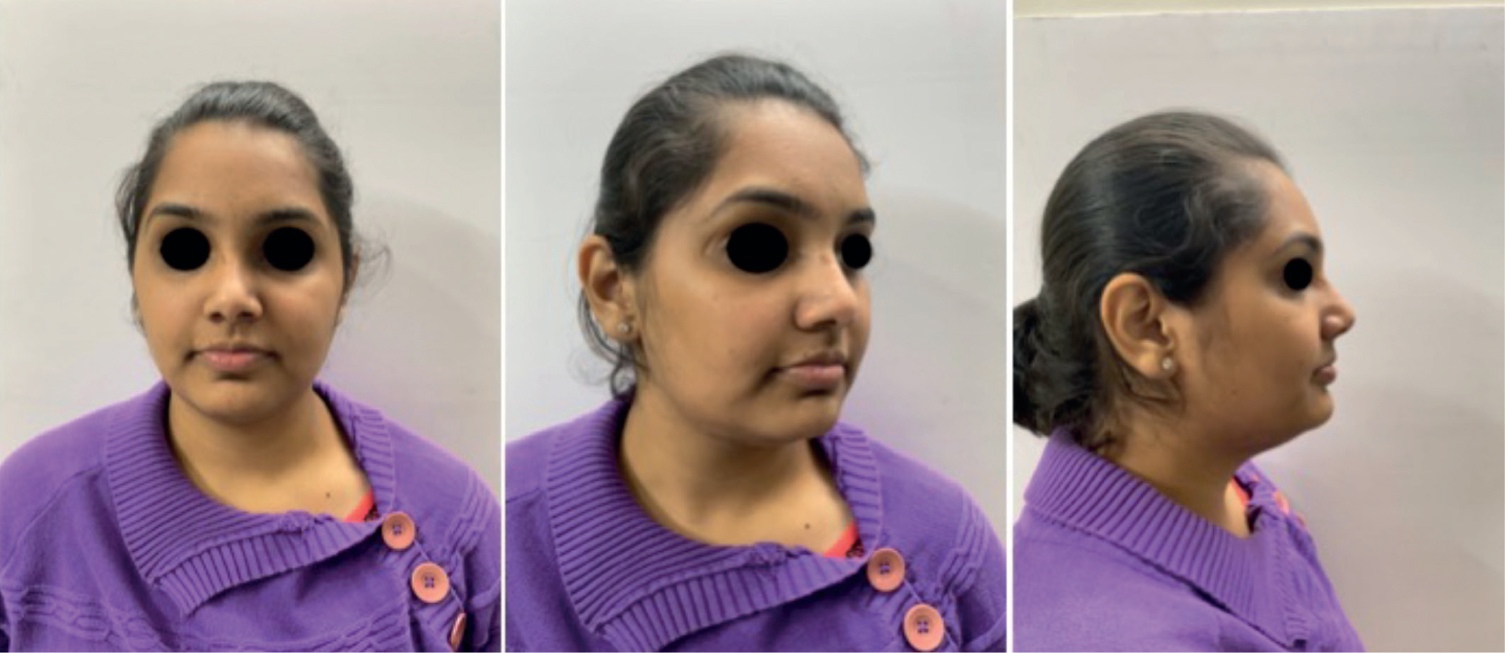
Figure 1. 2D views of a patient.

3D photogrammetry was suggested as a sophisticated procedure to create 3D models and maps. Photogrammetry has its origins during the First World War, where pilots gathered information from the combined use of photography and manned flights. The use of high-resolution cameras allows generation of similar models. The same has been applied in dentistry to create 3D models. This procedure can be expensive, but it is also technology friendly, and the evolution of mobile phones with high resolution cameras and suitable applications allows the same procedure to be used by all clinicians.1,2,3 This Technique Tip highlights the use of a freely available mobile app to create 3D models that can be helpful in creating a 3D model of a face. The app is free to access and easy to use on other plaforms, such as tablets and laptops.

Workflow for creating a 3D facial model:
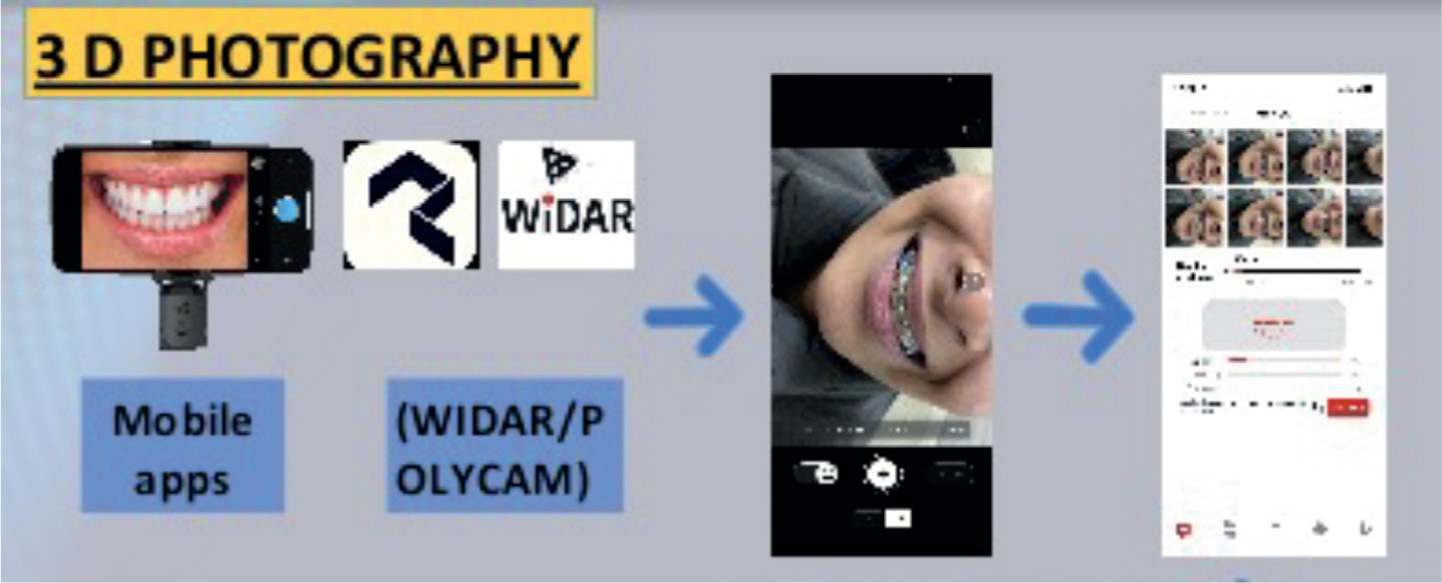
  • With a good internet connection, download the WIDAR/Polycam app from iOS/Android app store Figure 2);
  • Open the app and log in;
  • Hold the camera and tap the record button;
  • Move the phone around the face from side to side to record the facial features;
  • Images can be reviewed in the ‘Preview’ section;
  • A minimum of 20 images are recorded, uploaded and processed within the app;
  • The 3D image is generated and can be viewed in the app and downloaded to be shared for patient education and treatment planning (Figures 3 and 4).
  • Figure 2. Mobile apps and their use.
    Figure 3. 3D model generation and representative views of the same patient.
    Figure 4. 3D VTO of a patient.

    Conclusion

    Many features of the face, which may be missed in a 2D photograph, may be evaluated by the use of a 3D scanning app. These apps are also useful for patient education and will allow 3D photographs and VTO to become a part of routine clinical diagnosis.